
Explore chapters on mild and severe Ventriculomegaly, Intracranial hemorrage, Porencephaly, and Aqueductal stenosis.
Abnormal ventricles
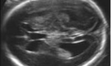
Mild Ventriculomegaly
There is marked biologic variability in the size of the fetal lateral ventricles. Empiric data suggests that fetuses with a width of the atrium (or posterior horn) of lateral ventricles of 10 mm or more have an increased risk of abnormal outcome. When the measurement is 10-15mm the risk is relatively low.
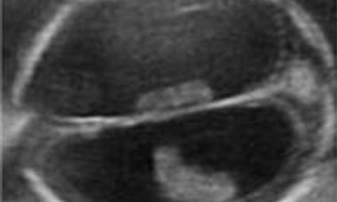
Severe Ventriculomegaly
The current definition is an increased size of the lateral ventricles, with a transverse diameter of the atrium in excess of 15mm without evidence of other cerebral malformations. It is a rare condition, usually a part of complex cerebral abnormalities, less frequently the consequence of obstructed cerebrospinal fluid turnover.
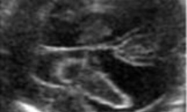
Intracranial Hemorrage
Intracranial hemorrage may occur within the lateral ventricles or in the subdural space. The sonographic appearance changes with time. An echogenic collection is first seen, and in the following days it develops into a complex mass frequently complicated by sever ventriculomegaly.
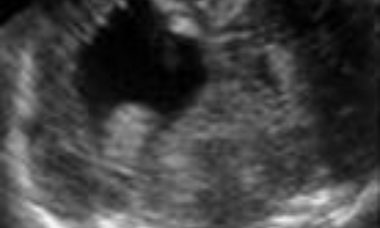
Porencephaly
Porencephaly is characterized by single or multiple cysts replacing the brain parenchyma. The cyst may communicate with the lateral ventricle, subarachnoid space or both. It is usually a sporadic condition caused by hemorrage, ischemia or infections.
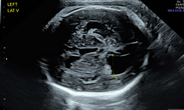
Aqueductal Stenosis
Aqueductal stenosis (AS) results from a narrowing or occlusion of the aqueduct of Sylvius.