
Explore chapters on choledocal cysts, mesenteric cysts, intenstinal duplication cysts, hepatic cysts, meconium pseudocyst, urinoma, splenic cysts and ovarian cysts
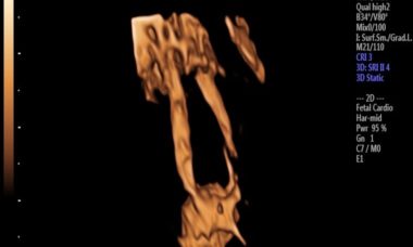
Choledochal Cysts
Congenital choledochal cysts are characterised by cystic or fusiform dilatation of the extra- and intrahepatic biliary system. The incidence of congenital choledochal cysts is rare in western population whilst it increases up to 1 in 10,000 in East Asian populations including Japan and China.
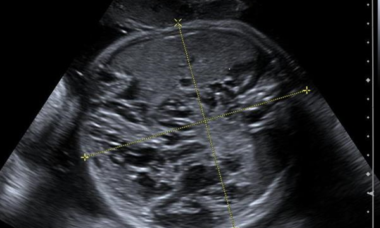
Mesenteric Cyst
A mesenteric cyst is suggested when an avascular, thin-walled, anechoic midline cystic intraabdominal mass is identified in the fetus. These cysts can be uni- or multilocular, and of variable size and multilocular cysts can extend to the abdominal wall, buttocks, or fetal legs. They are thought to arise from obstructed lymphatic drainage.
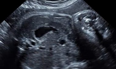
Intestinal Duplication Cyst
Intestinal duplication cysts are embryonic abnormalities where extra intestinal tissue is present adjacent to normal bowel. Although most are typically small bowel in origin, they can arise from anywhere along the gastrointestinal tract. In most cases, the extra intestinal tissue is closed at both ends.
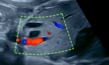
Congenital Hepatic Cyst
Congenital hepatic cysts are rare hepatobiliary malformations which typically present as a single, unilocular upper abdominal cystic mass in the fetus.
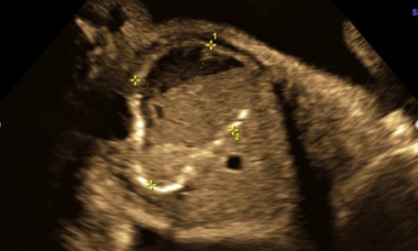
Meconium Pseudocyst
A meconium pseudocyst presents as a complication of meconium peritonitis in which meconium-filled bowel dilates to form a mass proximal to a site of intestinal obstruction or between atretic or stenotic small bowel segments. Meconium pseudocyst is differentiated from cystic type meconium peritonitis by a continuous smooth muscle layer between the cyst and the normal intestine with loss of the mucosal epithelium, as well as by communication between the cyst and the proximal dilated bowel.
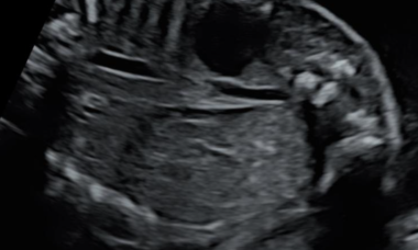
Urinoma
Fetal urinomas occur as a result of rupture of the renal collecting system which results in a hypoechoic elliptical or crescent-shaped fluid collection surrounding or adjacent to the kidney and within the peri-renal fascia.
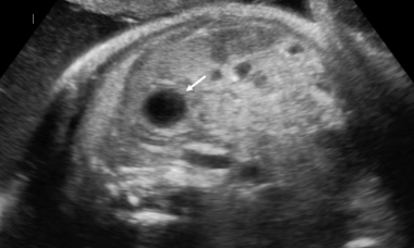
Congenital Splenic Cyst
Congenital splenic cysts (CSCs) are fluid-filled lesions located within the splenic parenchyma. There are often small and almost always asymptomatic. However, occasionally they can be large enough to be seen protruding from the internal surface of the spleen and can compress neighboring viscera.
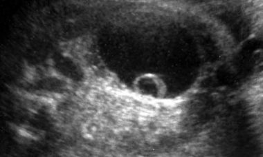
Fetal Ovarian Cysts
Fetal ovarian cysts are fluid filled masses arising from the fetal ovary, measuring at least 20 mm in diameter. They are thought to develop from fetal exposure to maternal and placental hormones.