
Tuberous Sclerosis is a neurocutaneous disorder with a hereditary component affecting diverse organ systems. The incidence is around 1:5 000-10 000 births.
Tuberosus Sclerosis with cortical hamartomas
Abstract: Tuberous Sclerosis is a neurocutaneous disorder with a hereditary component affecting diverse organ systems. The incidence is around 1:5 000-10 000 births. Cerebral hamartomas or cortical tubers are common manifestations and these lesions. They are composed of disorganized but mature cells, mostly a combination of neuronal or ganglion cells, glial cells and blood vessels which can have deleterious effects on the patient's health. This pathology is associated with genetic mutations of TSC gene.
Authors: Esteban Lizárraga-Cepeda1, Andrea G Carrillo-Obregón1, Aldo M Cruz-Hernández1, Gerardo Sepúlveda-González1, Tayde Arroyo-Lemarroy1, Noe E Nava-Guerrero1
- Hospital Regional Materno Infantil de Nuevo León, México, Departamento de Medicina Materno Fetal Institution/ Department
Keywords: Tuberosus Sclerosis, hamartomas, cortical hamartomas, cortical tubers, Gene mutations in TSC1 or TSC2, Bourneville disease.
Reviewers: Karen Fung-Kee-Fung
View the Patient Information leaflet
Definition
Tuberous Sclerosis (TSC) is a neurocutaneous disorder affecting diverse organ systems leading to brain, ocular, cardiac, pulmonary, hepatic, renal, and skin hamartomas. The term tuberous describes the potato-like consistency of gyri with hypertrophic sclerosis. (1,2)
Tuberous sclerosis is characterized by the development of benign brain tumors in multiple locations, including subependymal nodules, cortical tubers, and giant cell astrocytomas, with diverse clinical manifestations such as intractable focal epilepsy and neuropsychiatric disorders occurring throughout development, during infancy and even in adulthood. (2,3) Cerebral hamartomas are defined as lesions composed of disorganized but mature cells, mostly a combination of neuronal or ganglion cells, glial cells, and blood vessels.
ICD code
Q85.1 Tuberous sclerosis. (4)
Incidence
1:5,000-10,000 live births. (5-7)
Etiology
There is no elucidated etiology, but a “two-hit” theory has been proposed. In patients with tuberous sclerosis, all the somatic cells have a germline mutation (first hit) in one allele of either the TSC1 or TSC2 gene, which can cause haploinsufficiency of the TSC1/TSC2 complex. When an additional somatic mutation (second hit) occurs during mitosis, the function of the TSC1/TSC2 complex becomes null. (8) Some studies (9,10) have demonstrated that TSC-associated tumors (hamartomas) could occur due to the two-hit theory (11). The second hit is usually a small deletion of either TSC1 or TSC2, causing loss of heterozygosity. The incidence of this is reportedly low in brain tumors. (8)
Pathogenesis
Up to 85% of the cases are due to an autosomal dominant disorder due to variants in TSC1 or TSC2 genes (7,8,12-14), which are believed to function as tumor suppressor genes if they function correctly; or by overactivation of the mTOR pathway, which favors the formation of benign tumors in different organs and systems. (1,2 y 7) Hamartin is a protein encoded by the TSC1 gene on chromosome 9q34, which, in conjunction with the tuberin protein encoded by the TSC2 gene on chromosome 16p13.3, forms a complex. (12,15) Hamartin plays a crucial role in helping to stabilize the tuberin, allowing it to function correctly (16). When one of the two proteins (hamartin or tuberin) is not correctly codified, is deficient, or is not present, this complex is not formed and cannot exert its inhibitory function of the mTOR system. Likewise, tuberin is essential for normal brain development and the proper formation and differentiation of the cardiomyocyte. (17,18)
The C-terminus of tuberin has extensive sequence homology to the catalytic domain of GTPase-activating protein 3 (GAP3) that stimulates its intrinsic activity. These, in turn, cleave a phosphate group from GTP to form GDP, thereby inactivating GTP-binding proteins such as ras and the ras-like proteins Rap-1A and rab5a. Ras is one of the families of proteins that controls the cell cycle. (1,19,20) Tuberin stimulates the GTPase activity of Rap-1A. Since there is a deficiency of tuberin-hamartin in TSC, there is a lack of GTPase production and its function; therefore, there is no adequate regulation of the cell cycle. Cells spend less time in G1 (the resting phase of the cell cycle), and quiescent cells are induced to enter the cell cycle. (19)
The mTOR system is responsible for some aspects of cell proliferation. When there is a regulation failure in the mTOR system, there will be hyperstimulation of the proliferation system (21), thus favoring changes in cell migration (22), glucose uptake and metabolism (23), and angiogenesis (24), resulting in the generation of tumours and dysgenesis, leading to hamartomas at the cortical level.
Abnormal neuronal development likely plays a role in the neurologic manifestations. The alterations and tumours at the cortical level secondary to this lack of protein regulation will be responsible for brain dysfunction during intrauterine life and at birth. (8,21) The severity of the condition will depend on the location and size of the lesions. De-novo variants account for approximately two-thirds of cases; heterozygous germline and mosaic mutations have been reported. (2) There is a small portion of patients with cortical hamartomas in which there will be no evidence of alteration in the TSC1 and TSC2 genes; the etiology of these cases is still unknown. Of note, the altered portion at the genetic level of TSC1-TSC2 will not necessarily be related to the severity of the clinical presentation.
Pathology
The primary neuropathological lesions in the brain of patients affected by TSC are cortical tubers, subependymal nodules, and subependymal giant cell astrocytomas (SEGAs). Cortical tubers are observed in 80–90% of patients with TSC; it consists of areas of cortical disorganization containing different cell types, including dysmorphic neurons, giant cells, and reactive astrocytes (“balloon cells”). (2, 25) Subependymal nodules are considered the precursor lesions of SEGAs. According to the World Health Organization (WHO) classification of tumours of the central nervous system (CNS), SEGAs represent benign and slow-growing tumors composed of large ganglion-like astrocytes with a mixed glio-neuronal feature and correspond histologically to WHO grade 1. (2)
Associated anomalies
In utero features: Ventriculomegaly (26), cardiac fibromas, cardiac abnormalities and arrhythmias, renal cysts, and fetal hydrops. (7)
Post-natal features: Retinal and non-retinal abnormalities, lymphangioleiomyomatosis, renal cell carcinoma, coarctation of the aorta, hypopigmented macules. (1)
Recurrence risk
Autosomal dominant recurrence risk of 50% if one of the parents is affected. De novo mutations have no increased risk, comprising about 65% of cases. (7,12)
Diagnosis
Prenatal detection of this condition is rare. (27). When there is clinical suspicion, an ultrasound assessment should be performed, or an intentional workup should be carried out in patients with a genetic history of hamartomas using new genome sequencing techniques. Neurosonography should also be considered in the evaluation of these cases. Genetic and clinical diagnostic criteria for tuberous sclerosis complex were previously established. These criteria include 11 major and seven minor features (2). The definite diagnosis is made with two major features or one major feature with two minor features. (28)
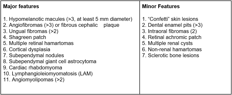
However, identifying these manifestations is limited during pregnancy because only a few of these criteria can be identified in utero but are easily assessed after birth. (25) Amniocentesis will facilitate molecular testing on a fetus when TS is suspected. When a family mutation is known, a chorionic villous biopsy is recommended. It is also possible to perform a preimplantation genetic study to select embryos not affected by the disease when the family mutation is known. Molecular testing of the TSC1 and TSC2 genes yields a positive mutation result for 75-90% of TSC-affected individuals, making identifying a pathogenic mutation an independent diagnostic criterion, regardless of the clinical findings. (2) Approximately 70% to 80% of individuals who meet definite diagnostic criteria have a small identifiable TSC1 or TSC2 gene mutation. The remaining individuals probably have large gene deletions, somatic mosaic mutations, and mutations in unanalyzed gene noncoding regions rather than an additional TSC gene locus. (12) Of note, a negative genetic test does not exclude the diagnosis because it may be due to mosaicism. Early genetic testing is essential for counseling and managing patients with tuberous sclerosis complex and their families, including prenatal assessments for families with a known affected member. Early diagnosis can lead to appropriate care, starting with improved potential for disease modification. (2,28) Advances in next-generation sequencing have made detecting mutations in the TSC1 and TSC2 genes more accurate and accessible, and pathogenic variants are now detectable in more than 90% of affected individuals. (2,29)
Differential diagnosis
Other central nervous system tumors such as ganglioglioma, oligodendroglioma, or low-grade astrocytoma should also be ruled out. (26)
Implications for sonographic diagnosis
Imaging tools such as fetal neurosonography and fetal MRI (FLAIR preferred), have shed new light on antenatal cerebral lesions of TSC. These imaging studies indicate that cortical tubers and subependymal nodules are the most common lesions detected prenatally in the early gestational period, usually between 10-20 weeks. (28)
An accurate diagnosis of the spectrum of fetal brain lesions included in TSC is feasible by neurosonography, and the findings are similar to those obtained by MRI. (30) Cardiac rhabdomyomas are the most common fetal cardiac tumours and the most common prenatal sign of TSC. The earliest antenatal sonographic detection of this cardiac tumour was reported at 15 weeks, whereas most cases are described after 24 weeks of gestation. Sonographically, cardiac rhabdomyomas appear as round, homogenous, hyperechogenic, intramural, or intracavitary masses, sometimes multiple, usually located in the ventricles and septal wall and occasionally in the atrium or pericardium. (28)
Implications for sonographic screening
Currently, there is no evidence in the literature regarding screening for the detection of this condition. Nonetheless, associated findings have been described since week 15, but those findings are more commonly observed in the middle of the second trimester. Considering prior evidence, routine evaluation during the second trimester with a structural ultrasound between weeks 18-22 is suggested, and a third-trimester assessment should also be considered. (31)
Prognosis
Tumour size, the occurrence of fetal dysrhythmias, such as ectopic beats, supraventricular tachycardia, or bradycardia, and the development of fetal hydrops are considered strong predictors of a negative neonatal outcome. (24) The correct identification and measurement of the brain lesions are essential because there is an association between the number and size of the tubers and impaired neurodevelopmental outcomes. (32)
Management
Presently there is no fetal therapy available. Nevertheless, close follow-up with ultrasound scans every four weeks is prudent to monitor the evolution of neurologic and cardiac tumours and screen for the development of arrhythmias, hydrocephalus, or fetal hydrops. (7) Delivery at a hospital with a neonatal intensive care unit at ~38 weeks of gestation is indicated if there are no fetal or maternal complications. (7) There is no contraindication to vaginal delivery. Cesarean section is generally preferred if there is fetal hydrops. (7) Multidisciplinary team will be required to comprehensively assess the newborn status and provide ongoing care after delivery.
References
1. Curatolo P, Bombardieri R, Jozwiak S. Tuberous sclerosis. Lancet 2008; 372:657.
2. Curatolo P, Specchio N, Aronica E. Advances in the genetics and neuropathology of tuberous sclerosis complex: edging closer to targeted therapy. Lancet Neurol 2022; 21:843.
3. Diehl B, Prayson R., Najm I., Ruggieri P. Hamartomas and epilepsy: clinical and imaging characteristics. Seizure 2003; 12: 307–311.
4. https://icd.who.int/browse10/2010/en#/Q85.1
5. Northrup H, Koenig MK, Pearson DA, Au KS. Tuberous sclerosis complex. In: GeneReviews [Internet], Adam MP, Ardinger HH, Pagon RA, et al. (Eds), University of Washington, Seattle 2020. Available at: https://www.ncbi.nlm.nih.gov/books/NBK1220/
6. Hallett L, Foster T, Liu Z, et al. Burden of disease and unmet needs in tuberous sclerosis complex with neurological manifestations: systematic review. Curr Med Res Opin 2011; 27:1571.
7. Nicolaides K., Cruz J., Hayashi N., Molina F., Litwińska M. and Vichos D. Fetal abnormalities-Abnormalities by system, brain, Tuberous sclerosis. https://fetalmedicine.org/education/fetal-abnormalities/brain/tuberous-sclerosis
8. Mizuguchi, M.; Ohsawa,M.; Kashii, H.; Sato, A. Brain.Symptoms of Tuberous Sclerosis Complex: Pathogenesis and Treatment. Int. J. Mol. Sci. 2021, 22, 6677. https://doi.org/10.3390/ijms22136677
9. Green, A.J.; Smith, M.; Yates, J.R. Loss of heterozygosity on chromosome 16p13.3 in hamartomas from tuberous sclerosis patients. Nat. Genet. 1994, 6, 193–196.
10. Henske, E.P.; Scheithauer, B.W.; Short, M.P.; Wollmann, R.; Nahmias, J.; Hornigold, N.; Slegtenhorst, M.; Welsh, C.T.; Kwiatkowski,D.J. Allelic loss is frequent in tuberous sclerosis kidney lesions but rare in brain lesions. Am. J. Hum. Genet. 1996, 59, 400–406.
11. Niida, Y.; Stemmer-Rachamimov, A.O.; Logrip, M.; Tapon, D.; Perez, R.; Kwiatkowski, D.J.; Sims, K.; MacCollin, M.; Louis, D.N.; Ramesh, V. Survey of somatic mutations in tuberous sclerosis complex (TSC) hamartomas suggests different genetic mechanisms for pathogenesis of TSC lesions. Am. J. Hum. Genet. 2001, 69, 493–503.
12. Au, K. S., Williams, A. T., Roach, E. S., Batchelor, L., Sparagana, S. P., Delgado, M. R., Wheless, J. W., Baumgartner, J. E., Roa, B. B., Wilson, C. M., Smith-Knuppel, T. K., Cheung, M. Y. C., Whittemore, V. H., King, T. M. & Northrup, H. (2007, febrero). Genotype/phenotype correlation in 325 individuals referred for a diagnosis of tuberous sclerosis complex in the United States. Genetics in Medicine, 9(2), 88-100. https://doi.org/10.1097/gim.0b013e31803068c7
13. European Chromosome 16 Tuberous Sclerosis Consortium. Identification and characterization of the tuberous sclerosis gene on chromosome 16. Cell 1993; 75:1305.
14. van Slegtenhorst, M.; Nellist, M.; Nagelkerken, B.; Cheadle, J.; Snell, R.; van den Ouweland, A.; Reuser, A.; Sampson, J.; Halley, D.; van der Sluijs, P. Interaction between hamartin and tuberin, the TSC1 and TSC2 gene products. Hum. Mol. Genet. 1998, 7, 1053–1057.
15. Rosset C, Netto CBO, Ashton-Prolla P. TSC1 and TSC2 gene mutations and their implications for treatment in tuberous sclerosis complex: a review. Genet Mol Biol. 2017;40(1):69–79.
16. van Slegtenhorst M, de Hoogt R, Hermans C, et al. Identification of the tuberous sclerosis gene TSC1 on chromosome 9q34. Science 1997; 277:805.
17. Rennebeck G, Kleymenova EV, Anderson R, Yeung RS, Artzt K, Walker CL. Loss of function of the tuberous sclerosis 2 tumor suppressor gene results in embryonic lethality characterized by disrupted neuroepithelial growth and development. Proc Natl Acad Sci U S A. 1998 Dec 22;95(26):15629-34. doi: 10.1073/pnas.95.26.15629. PMID: 9861021; PMCID: PMC28095.
18. Salussolia CL, Klonowska K, Kwiatkowski DJ, Sahin M. Genetic etiologies,diagnosis, and treatment of tuberous sclerosis complex. Annu Rev Genomics Hum Genet. 2019;20:217–40.
19. Soucek T, Pusch O, Wienecke R, et al. Role of the tuberous sclerosis gene-2 product in cell cycle control. Loss of the tuberous sclerosis gene-2 induces quiescent cells to enter S phase. J Biol Chem 1997; 272:29301.
20. Wienecke R, König A, DeClue JE. Identification of tuberin, the tuberous sclerosis-2 product. Tuberin possesses specific Rap1GAP activity. J Biol Chem 1995; 270:16409.
21. Hengstschläger, M.; Rodman, D.M.; Miloloza, A.; Hengstschläger-Ottnad, E.; Rosner, M.; Kubista, M. Tuberous sclerosis gene products in proliferation control. Mutat. Res. 2001, 488, 233–239.
22. Mizuguchi, M.; Yamanouchi, H.; Becker, L.E.; Itoh, M.; Takashima, S. Doublecortin immunoreactivity in giant cells of tuberous sclerosis and focal cortical dysplasia. Acta Neuropathol. 2002, 104, 418–424.
23. Jiang, X.; Kenerson, H.; Aicher, L.; Miyaoka, R.; Eary, J.; Bissler, J.; Yeung, R.S. The tuberous sclerosis complex regulates trafficking of glucose transporters and glucose uptake. Am. J. Pathol. 2008, 172, 1748–1756.
24. Brugarolas, J.; Kaelin, W.G., Jr. Dysregulation of HIF and VEGF is a unifying feature of the familial hamartoma syndromes. Cancer Cell 2004, 6, 7–10.
25. Mongrain et al. A case report of severe tuberous sclerosis complex detected in utero and linked to a novel duplication in the TSC2 gene. BMC Neurology (2020) 20:324 ://doi.org/10.1186/s12883-020-01905-yhttps
26. DiPaolo D, Zimmerman RA. Solitary cortical tubers. AJNR Am J Neuroradiol. 1995 Jun-Jul;16(6):1360-4. PMID: 7677041; PMCID: PMC8337854.
27. M. Sgro, T. Barozzino, A. Toi, J. Johnson,M. Sermer, D. ChitayatPrenatal detection of cerebral lesions in a fetus with tuberous sclerosis. Ultrasound Obstet Gynecol 1999;14:356–359
28. Dragoumi P, O’Callaghan F, Zafeiriou DI. Diagnosis of tuberous sclerosis complex in the fetus. Eur J Paediatr Neurol 2018; 22: 1027–34.
29. Peron A, Au KS, Northrup H. Genetics, genomics, and genotypephenotype correlations of TSC: Insights for clinical practice. Am J Med Genet C Semin Med Genet 2018; 178: 281–90
30. F. Correa1, B. Puerto2, G. Quiroz3, M. Forteza4, F. Vinals ̃. Neurosonographic findings in fetal tuberous sclerosis complex. Ultrasound in Obstetrics & Gynecology 2017; 50 (Suppl. 1): 154–256.
31. Salomon, L.J., Alfirevic, Z., Berghella, V., Bilardo, C.M., Chalouhi, G.E., Da Silva Costa, F., Hernandez-Andrade, E., Malinger, G., Munoz, H., Paladini, D., Prefumo, F., Sotiriadis, A., Toi, A. and Lee, W. (2022), ISUOG Practice Guidelines (updated): performance of the routine mid-trimester fetal ultrasound scan. Ultrasound Obstet Gynecol, 59: 840-856. https://doi.org/10.1002/uog.24888
32. M. Sanz-Cortes, J. M. Martinez, M. Bennasar, B. Puerto E. Gratacos Prenatal diagnosis of tuberous sclerosis and analysis using magnetic resonance spectroscopy. Ultrasound Obstet Gynecol 2010; 36: 521–524.
This article should be cited as: Lizárraga-Cepeda E, Carrillo-Obregón A, Cruz-Hernández A, Sepúlveda-González G, Arroyo-Lemarroy T, Nava-Guerrero N: Tuberosus Sclerosis with cortical hamartomas, Visual Encyclopedia of Ultrasound in Obstetrics and Gynecology. www.isuog.org, January 2023.
Leave feedback or submit an image
We rely on your feedback to update and improve VISUOG. Please use the form below to submit any comments or feedback you have on this chapter.
If you have any images that you think would make a good addition to this chapter, please also submit them below - you will be fully credited for all images used.
Feedback form
Please note that the maximum upload size is 5MB, and larger images and video clips can be sent to [email protected].