
The presence of an extra limb in a fetus is a rare but important congenital anomaly. These limbs are usually not functional but involve significant dysmorphology and if uncorrected, may lead to major quality of life issues.
Supernumerary Limb
Abstract: The presence of an extra limb in a fetus is a rare but important congenital anomaly. These limbs are usually not functional but involve significant dysmorphology and if uncorrected, may lead to major quality of life issues. The correction of these anomalies depends on the site and size of the extra limb and the nature of its attachment to normal body tissues. Appropriate prenatal diagnosis and a plan for postnatal management can help these fetuses achieve a good quality of life postnatally.
Authors: Chinmayee Ratha1, Karra Sahithi Reddy1, Vidya Sudha Panja1
- Resolution Fetal Medicine Centre and Research Institute, Secunderabad, Telangana,India
Reviewer: Asma Khalil, Karen Fung-Kee-Fung
View the Patient Information sheet
Definition
Fetal supernumerary limb or “polymelia” is a rare congenital malformation characterized by presence of additional limb elements apart from normally attached limbs. These extra limbs may be attached to various parts of the fetal body and based on this they can be classified as: Cephalomelia- attached to the fetal head, thoracomelia- attached to the thorax, pygomelia - attached to the pelvis or notomelia- which has an attachment to the region of the embryological notochord, which contributes to the development of the axial skeleton and eventually becomes the nucleus pulposus of the intervertebral disc.1
ICD Code
Q 79.9 congenital deformity NOS
Incidence
Isolated limb duplication is a rare congenital condition and only a few cases have been documented in literature. Interestingly, in a review article in 2011, Klassen et al 2have cited references to cases of polymelia that have been documented since the early nineteenth century. In some papers, “polydactyly” has also been referred as a partial limb duplication. Overall, there is a lack of robust definition and classification of extra limbs and thus the exact incidence remains unknown. Currently, there are no “case series” or larger cohorts reported as yet hence it is assumed that the incidence of supernumerary limbs remains very rare and sporadic.
Pathogenesis
Limb differentiation occurs usually between the 4th and 5th weeks of embryonic development with two pairs of primitive limb buds - anterior and posterior, that project from either side of the developing embryo. The pattern of development continues along 3 axes in “dorsoventral”, “proximodistal” and “anteroposterior”.
The differentiating regions are comprised of cells from two germ layers– the ectoderm and mesoderm and research in cell signaling pathways has started to unravel the role of local cytokines in the co-ordinated flow of events in normal limb development. Three important signaling centres have been identified as the apical ectodermal ridge(AER) , zone of proliferating activity(ZPA) and the dorsal ectoderm3. Hence it may be extrapolated that any disruption or deviation from the expected developmental process can lead to limb malformations. Lin et al 4have proposed a theory of “cross talk” between signalling factors like the fibroblast growth factors (FGF), Bone morphogenetic proteins(BMP), Engrailed- 1(En-1) etc5. The primordial limb buds can be affected by multiple signalling pathways and in any situation where the apical ectodermal ridge is transplanted, an ectopic, extra limb development can ensue. Some authors have termed the process as “excess of morphogens” 6during the development process can lead to polymelia as shown in Fig 1. In developmental terms, supernumerary limbs would be classified either “malformation” or “deformation” sequence. Some authors have also postulated the possibility of incomplete separation of monozygotic twins5 leading to appearance of extra limbs or polymelia.
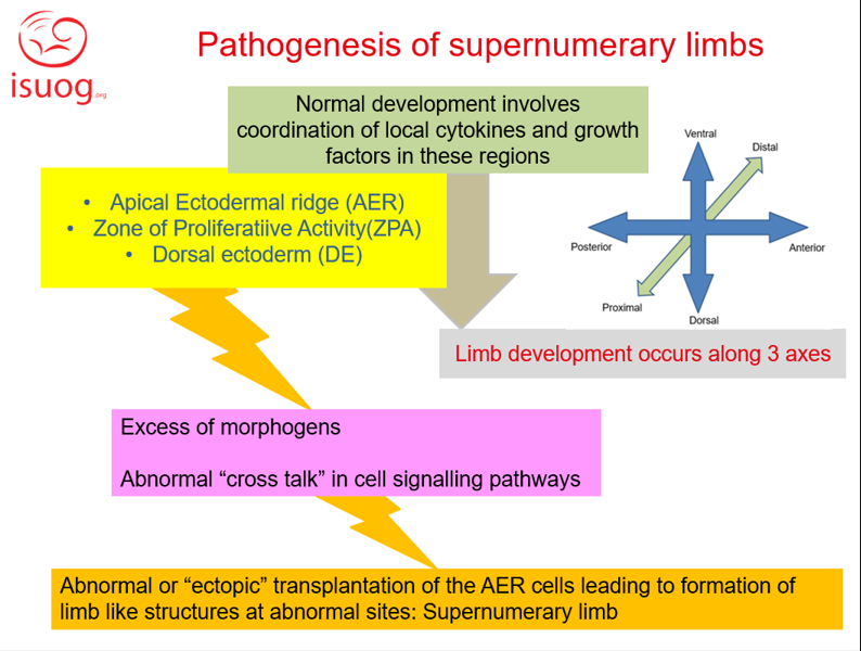
Figure 1: Pathogenesis of supernumerary limb
Etiology
Multiple adverse embryogenic influences could be responsible for development of supernumerary limbs in a fetus. The generic etiological factors for these adverse effects could be many like:
- Single gene disorders (e.g HOX, T-Box genes)7
- Chromosomal abnormalities (e.g 22q11 microdeletion)2
- Vascular events8
- Maternal exposure to teratogens (e.g retinoids, pesticides etc)2
- Unknown causes
Pathology
In the process of limb differentiation during embryonic development in vertebrates it has been suggested that a process called "specification” instructs the cells of the future limb regarding the type of tissue they should form. The human limb duplication results from an error in this process.
The sequence of specification goes from proximal to distal sites and based on this the classification of human limb duplication was proposed:
a) Specification defect- complete absence of bones and related soft tissue or a supernumerary limb
b) Post specification defect- diminished or absent bone structures with relatively normal soft tissue
This concept of specification also helps us to time the occurrence of teratogenic event – the more distal the duplication, the later the teratogenic event must have affected the normal process of limb development.
Literature has also revealed that vascular deformity is very often associated with limb deformity.
The supernumerary limb can affect cosmetic appearance externally. Polymelia can also limit movement and postural comfort of the baby based on its position. Depending on its attachment to internal structures, functional problems can be present– eg. if it is connected to the spine, neuromotor disturbance is possible.
Due to the above mentioned factors, it becomes imperative to excise the extra limb and correct the physical deformity with an aim to restore normal function of the body and minimize discomfort.
Associated anomalies
Spinal cord anomalies, OEIS (Omphalocoele, exstrophy of cloaca, imperforate anus, spinal defects) complex, renal abnormalities.
Recurrence risk
The recurrence risk of this anomaly is unknown but thought to be exceedingly rare as only few cases are documented to date. A genetic predisposition has been speculated in one case report from Niger8 where the author has cited 3 similar cases in 3 different couples – all of whom were consanguineous parents belonging to a closely knit ethnic group.
Diagnosis
Polymelia is a diagnosis made on the basis of presence of an extra limb in the fetus in addition to its normal limbs. In this case the exact diagnosis was “notomelia” as the extra limb element was attached to the duramater of the fetal spine. There was however no open spinal dysraphism associated in our case.
Differential diagnosis
Parasitic twin, Spinal Dysraphism, Sacrococcygeal teratoma
Table 1: Summary of major differential diagnoses for fetal supernumerary limb
In our illustrated case the possibility of sacrococcygeal teratoma was eliminated by the position of the mass in the lumbar region, somewhat laterally placed, with bony elements and minimal vascularity. There was no involvement of the spine and open spinal dysraphism was unlikely.
We could not rule out the possibility of a “parasitic twin” antenatally and kept it as a close differential diagnosis to notomelia. The antenatal care plan did not get affected with this lack of clarity as the final management plan would involve separation surgery in either situation.
Implications for sonographic diagnosis
A standardized and detailed antenatal ultrasound examination is imperative to accurate diagnosis, and elimination of other differential diagnoses and contribute to the successful outcome of cases of supernumerary limb. After evaluation of the normal fetal limbs the site of the attachment of the extra appendage to the fetal body will help classifiy the supernumerary limb – it may be attached either to the fetal head (cephalomelia), spine (notomelia), pelvis(pygomelia) or thorax(thoracomelia). During this ultrasound examination it is important to rule out spinal dysraphism by visualizing the entire extent of the fetal spine in longitudinal, coronal and axial views ruling out any canal dilatation, tethering of the fetal spinal cord or distortion in the arrangement of the vertebrae.
A sacrococcygeal teratoma (SCT) will appear as a heterogenous mass attached to the sacral spine with obvious vascular connections and possible distortion of the sacral spine. The structure is usually globular in shape and does not exhibit a usual limb configuration. The differential diagnosis of a parasitic twin has to be always considered in cases of supernumerary limb as they may be indistinguishable on antenatal ultrasound.
Antenatal counselling is based on ultrasound diagnosis and the final outcome depends on the correct correlation of antenatal and postnatal findings.
Implications for sonographic screening
Screening occurs at the time of routine anomaly scan. A systematic evaluation of the normal limbs should reveal all three segments of all four limbs with demonstration of normal movements at the joints as expected. If, in addition to these structures, extra limbs or limb segments are noted attached to any part of the fetal body, then the diagnosis of supernumerary limb is made. The spine should be carefully evaluated for any evidence of spinal dysraphism.
Prognosis
The prognosis of this kind of anomaly will depend on multiple factors, including etiology and associated anomalies. In cases of genetic syndromes and chromosomal disorders the prognosis may be guarded and quality of life may be affected with multisystemic involvement. In cases of an isolated polymelia, with no involvement of any other system that could result in motor and neuronal deficit postnatally, the prognosis is likely to be favourable. In most cases, postnatal surgical correction is possible.
In the absence of antenatal diagnosis, birth injury to both mother and fetus may be encountered, due to malpresentations or labour dystocia. Fortunately, In this age of antenatal ultrasound screening, this situation would be expected to be uncommon.
Management
Supernumerary limbs are generally surgically correctable. A standard ultrasound evaluation is a must for correct diagnosis. Detailed multidisciplinary counseling of the family is imperative, involving a specialist team that includes a fetal medicine and imaging expert, pediatric surgeon, and primary obstetrician is essential for planning for delivery and postnatal care. Timely detection and proper referral to the fetal medicine service plays a key role in every antenatally diagnosed fetal abnormality. Effective multidisciplinary communication with the parents of the affected fetus helps in optimizing the care pathway in such cases where postnatal surgical correction has to be planned.10
Prevention
Supernumerary limbs are very rare malformations in humans. Exposure to environmental teratogens like retinoids and pesticides can be avoided to prevent the occurrence of these anomalies. However, in most cases the etiology is unknown or related to genetic or chromosomal disorders for which there is currently no effective primary prevention.
References
1. Verma S, Khanna M, Tripathi VN, Yadav NC. Occurrence of polymelia in a female child. J Clin Imaging Sci. 2013 Apr 30;3:18. doi: 10.4103/2156-7514.111235. PMID: 23814690; PMCID: PMC3690670.
2. Klaassen, Z., Shoja, M. M., Tubbs, R. S., & Loukas, M. (2011). Supernumerary and absent limbs and digits of the lower limb: A review of the literature. Clinical Anatomy, 24(5), 570–575. doi:10.1002/ca.21102
3. Duboc V, Logan MP. Building limb morphology through integration of signalling modules. Curr Opin Genet Dev. 2009 Oct;19(5):497-503. doi: 10.1016/j.gde.2009.07.002. Epub 2009 Sep 2. PMID: 19729297
4. Lin GH, Zhang L. Apical ectodermal ridge regulates three principal axes of the developing limb. J Zhejiang Univ Sci B. 2020;21(10):757-766. doi:10.1631/jzus.B2000285
5. Montalvo N, Redrobán L, Espín VH. Incomplete duplication of a lower extremity (polymelia): a case report. J Med Case Rep. 2014;8:184. Published 2014 Jun 12. doi:10.1186/1752-1947-8-184
6. Kher AS, Gahankari DR, Tambwekar SR, Doraiswamy A, Iyer S, Bharucha BA, et al. Supernumerary limbs: A case report of a rare congenital anomaly. Ann Plast Surg 1996;37:549 52.
7. Barham G, Clarke NM. Genetic regulation of embryological limb development with relation to congenital limb deformity in humans. J Child Orthop. 2008;2(1):1-9. doi:10.1007/s11832-008-0076-2
8. Kelani, A., Moumouni, H., Issa, A., Younsaa, H., Fokou, H., Sani, R., … Catala, M. (2017). Notomelia and related neural tube defects in a baby born in Niger: case report and literature review. Child’s Nervous System, 33(3), 529–534. doi:10.1007/s00381-017-3337-x
9. Sharma G, Mobin SS, Lypka M, Urata M. Heteropagus (parasitic) twins: a review. J Pediatr Surg. 2010 Dec;45(12):2454-63. doi: 10.1016/j.jpedsurg.2010.07.0026
10. Ratha C, Optimising Multidisciplinary Perinatal Care in Correctable Foetal Anomalies. Current Progress in Obstetrics & GynaecologyVolume-6;2021, Pg 217-231
The article should be cited as: Ratha C., Reddy K.S., Panja V.S., Supernumerary Limb, Visual Encyclopedia of Ultrasound in Obstetric and Gynecology, www.isuog.org, October 2022.
Leave feedback or submit an image
We rely on your feedback to update and improve VISUOG. Please use the form below to submit any comments or feedback you have on this chapter.
If you have any images that you think would make a good addition to this chapter, please also submit them below - you will be fully credited for all images used.
Feedback form
Please note that the maximum upload size is 5MB, and larger images and video clips can be sent to [email protected].