This leaflet is to help you understand what Ventriculomegaly is, what tests you need, and the implication of having been diagnosed with Ventriculomegaly for you, your baby and your family.
What is ventriculomegaly?
If a doctor or a sonographer tells you that your baby has enlarged lateral ventricles, this condition is known as ventriculomegaly. In the brain there are five interconnected fluid-filled cavities. These are called ventricles. The ventricles produce and are filled with cerebro-spinal fluid; from the ventricles the cerebro-spinal fluid circulates into the central spinal canal.
During a routine ultrasound investigation, the width of the posterior part (‘posterior horn’ or ‘atrium’) of the two lateral ventricles, situated on each side of the midline, is measured. These two parallel cavities are narrow anteriorly (towards the front of the body) and wider posteriorly (towards the back). The posterior horn of the lateral ventricles is clearly visible at ultrasound examination as a roughly triangular black area (because it is filled with fluid) having inside a white lump of tissue called the “choroid plexus." This is a lump of tiny vessels that produce the cerebro-spinal fluid. The width of the atrium of the lateral ventricles is normal up to 10 mm.
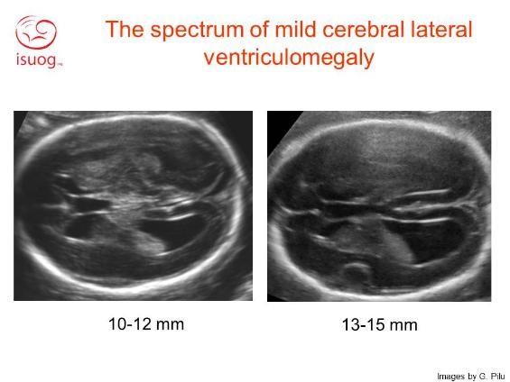
A width of 10-15 mm is called ventriculomegaly. Up to 12 mm we call it “mild” ventriculomegaly. If the posterior horn (atrium) measures more than 15 mm, it is known as hydrocephaly. It is not uncommon for lateral ventricles to be slightly larger in boys. Ventriculomegaly occurs in about 1% of fetuses.
How does ventriculomegaly happen?
There are a number of abnormal situations that can cause a blockage of the circulation of the cerebro-spinal fluid. A thin communication between the third and fourth ventricle, called the aqueduct, can become obstructed, leading to fluid accumulating above it and causing dilatation (widening) of the lateral ventricles. This is visible on ultrasound as enlarged fluid filled (black) spaces above the level of the obstruction.
Common causes of ventriculomegaly/hydrocephaly are:
-Infections
-Cerebral or spinal anomalies
-Chromosomal anomalies
-Bleeding in the brain
Should I have more tests done?
When the doctor or sonographer measures the lateral ventricles to be more than 10 mm wide, you will likely be offered a number of additional investigations:
- The fetus will be examined thoroughly to exclude other anomalies.
- Special attention will be given to anomalies of the brain and of the spine, as these can cause enlargement of the ventricles. The doctor or sonographer may suggest looking at the brain of the baby with a vaginal ultrasound scan.
- You will likely be offered an amniocentesis to look for problems in the number of chromosomes or large changes within the chromosomes. Chromosomes are where most of our genetic information is kept. We usually have 46 of them matched in pairs: 23 come from one parent and the other 23 come from the other parent. For example, people with Down syndrome have an extra chromosome number 21. Some fetuses with Down syndrome have enlarged ventricles, but there are also other genetic conditions associated with enlarged ventricles, for instance in male fetuses.
- Screening for infections to see if you have contracted an infection in pregnancy that may have caused the enlargement of the ventricles. Toxoplasmosis or CMV infection can cause an enlargement of the ventricles.
- In some cases, MR (magnetic resonance) imaging of the fetal brain may be offered later in pregnancy, to observe if the outer layer (cortex) of the brain is developing normally and try to better understand the reason behind the finding on ultrasound.
If all of these investigations are negative, your fetus is said to have an “isolated” ventriculomegaly.
The doctor will continue to follow your fetus with additional ultrasound scans to see whether the enlargement of the lateral ventricles remains stable, increases or decreases.
What does it mean for my baby after it is born?
The prognosis of ventriculomegaly will depend largely on whether an underlying cause has been determined. In cases of an “isolated” ventriculomegaly the prognosis is generally good. A mild enlargement of the ventricles may be normal in boys with a large head.
It is difficult to provide parents reliable figures regarding the chance of a neurological problem after birth in a baby diagnosed with ventriculomegaly during pregnancy. Although there are still insufficient data, the existing publications in the medical literature indicate that the risk of a neurological impairment is not higher than 10% when the ventriculomegaly is mild and isolated. This is about the same as the rest of the population.
If the ventricles have not increased in size during pregnancy and if no other explanations for the mild enlargement have been found, there is no specific indication for further investigations of the baby after birth. You or your doctor may however want to discuss this further.
Last update: March 2025
Download the leaflet: