
Chondroectodermal dysplasia (Ellis-Van Creveld syndrome) is a rare autosomal recessive congenital abnormality.
Chondroectodermal dysplasia
Abstract: Chondroectodermal dysplasia (Ellis-Van Creveld syndrome) is a rare autosomal recessive congenital abnormality. Prenatal diagnosis is based on the identification of moderately severe thoracic hypoplasia in addition with mild to moderate acromesomielia, postaxial polydactyly (which involves the hands in 100% of the cases), congenital heart disease (septum primum atrial defect in up to 50%) and ectodermal defects (dysplastic nails and teeth). Therefore, Ellis–Van Creveld syndrome can be suspected from prenatal ultrasound findings such as narrow chest, postaxial polydactyly of the hands and short limbs, and can also be confirmed on postnatal radiographic features.
Keywords: Chondroectodermal dysplasia, Ellis-Van Creveld syndrome, Polydactyly.
Synonyms: Ellis-Van Creveld Syndrome, mesoectodermal dysplasia, “six-fingered dwarfism”.
Authors: Enrique Gil-Guevara1; Olga Gómez del Rincón2
Department: 1.Instituto Peruano de Medicina y Cirugia Fetal / Lima, Peru
2.Hospital Clínic, Maternal-Fetal Medicine Department / Barcelona, Spain
View the Patient Information sheet
Chondroectodermal Dysplasia
Definition
Ellis-Van Creveld (EVC) syndrome, also called chondroectodermal dysplasia, mesoectodermal dysplasia, or chondrodystrophy, is a rare autosomal recessive congenital disorder of the skeletal dysplasia type.
ICD Code
Q77.6
OMIM # 225500
Incidence
There are approximately 150 cases reported worldwide1.
Pathogenesis
The name chondroectodermal dysplasia appears to be a generalized disorder in the maturation of endochondral ossification. Thus, the main features of this syndrome are both defective cartilaginous layer between the diaphysis and epiphysis and dysplasia of the ectodermal tissues (nails and teeth). Disorganized endochondral ossification is uniformly distributed and there is no lattice-advancing cartilage in the metaphysis. The following postnatal radiographic features are characteristic: shortened long bones with smooth rounded metaphysis, vertically short iliac bones, horizontally oriented acetabulum with medial and lateral spikes, short ribs, and normal vertebrae2,3.
Etiology
Is a genetic disorder caused by a mutation in the EVC gene located on the short arm of chromosome 4, in a region proximal to the gene involved in achondroplasia. Five different mutations have been associated with chondroectodermal dysplasia. Observation of inheritance patterns has illustrated that this is an autosomal recessive disease with variable phenotypic expression3.
Pathology
This syndrome is characterized by a variable spectrum of clinical findings, among the most common are chondrodystrophy, polydactyly, ectodermal dysplasia, and congenital cardiac anomalies. Oral manifestations of EVC syndrome include hyperplastic frenulum, absence of mucobuccal fold, gingival hypertrophy, hypodontia, dystrophic philtrum, and cleft palate4. The maxillary and mandibular alveolar processes may present notching or submucosal clefts.
Associated Anomalies
Congenital heart disease is the main associated anomaly, which affects up to 60% of cases (most commonly atrial septal defects, atrioventricular canal defect, ventricular septal defects, and hypoplastic left heart syndrome). Cleft palate is also seen in this disease. Central nervous system and urinary tract anomalies are also some of its rare associations4.
Recurrence Risk
The disease is inherited as an autosomal recessive trait, with a 25% risk of recurrence.
Diagnosis
Prenatal ultrasound features of chondroectodermal dysplasia are: mild to moderate acromesomelia (predominant shortening of the middle and distal segments of the limbs), moderate to severe thoracic hypoplasia with short ribs (narrow chest), postaxial polydactyly (which involves the hands in 100% of the cases and the feet in a significantly lower percentage, 10-25 % of cases) and congenital heart defects. Nevertheless, the cardiac anomaly most frequently associated with this syndrome are atrial septal defects, which are difficult to diagnose during fetal life due to the physiological patent foramen ovale5.
The ectodermal defects include dysplastic fingernails, neonatal teeth, partial anodontia, short upper lip bound by the frenulum to the alveolar ridge, and epispadias. Most of them are difficult to identify during prenatal ultrasound examination1,4,5.
Differential Diagnosis
The differential diagnosis of EVC includes Weyers syndrome, McKusick-Kaufman (MKK) syndrome, and Jeune syndrome. MKK syndrome, which is a recessively inherited disorder, can be distinguished from EVC by its characteristic presence of hydrometrocolpos. In Weyers syndrome, ectodermal disturbances resemble EVC, but unlike EVC, thoracic dysplasia, congenital heart defects, and dwarfism are absent. Jeune syndrome, an inherited form of dwarfism, is characterized by short limbs, similar to those observed in EVC. However, patients suffering from Jeune syndrome (asphyxiating thoracic dysplasia) do not exhibit fingernail hypoplasia, which is observed in patients with EVC (6). Short-rib polydactyly syndromes (SRPS) feature severe thoracic hypoplasia and micromelia, which are absent in EVC (Table 1).
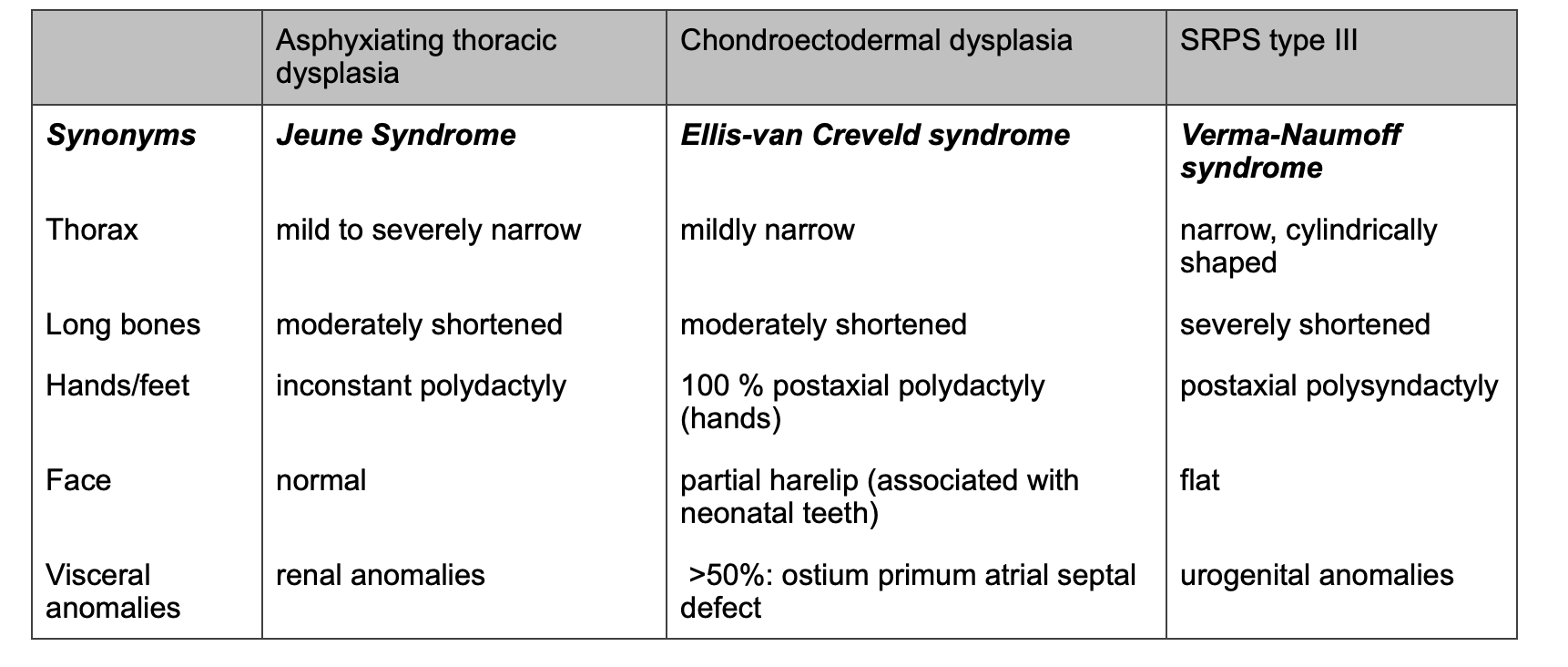
Table 1: Differential Diagnosis of Chondroectodermal Dysplasia.
Implications for sonographic diagnosis
In pregnancies at specific risk, a targeted vaginal first-trimester ultrasound may help to identify fetuses at risk since this is one of the severe skeletal dysplasias associated with increased nuchal translucency7. In case of potential recurrence, prenatal diagnosis using molecular genetic techniques is feasible from chorionic villus sampling (CVS).
Implications for sonographic screening
EVC is found in a high proportion of those fetuses where moderate thoracic hypoplasia is detected during a standard sonographic examination performed in the second trimester5. This finding should encourage evaluation of the fetal long bones, heart, fingers, and toes. Ostium primum atrial septal defect may be present in half of the cases.
Prognosis
The syndrome presents a 50% infant mortality rate, this is related to the severity of thoracic hypoplasia and consequent ventilatory problems. Survivors will have normal intelligence but short stature4,5. The cardiac anomaly can be successfully corrected. Dentition defects often require orthodontic procedures.
Management
Patients with EVC should be managed through a multidisciplinary approach. These patients require respiratory distress and heart failure treatment during the neonatal period.
Treatment should be started as early as possible to attain the best possible clinical outcome. Patients may require gingivectomy, frenectomy, and amputation of extra digits. Dental care after the eruption of teeth includes oral hygiene instructions, dietary counseling, and effective plaque control5.
Prenatal diagnosis of chondroectodermal dysplasia should encourage us to define the degree of thoracic hypoplasia to determine if it is a mild or a lethal case in which termination of pregnancy (TOP) can be offered before viability7. In the case of TOP, confirmation of the syndrome by autopsy is of high importance to future counseling.
References
- Howard, T.D., A.E. Guttmacher, W. McKinnon, M. Sharma, V.A. McKusick and E.W. Jabs, Autosomal dominant postaxial polydactyly, nail dystrophy, and dental abnormalities map to chromosome 4p16, in the region containing the Ellis-van Creveld syndrome locus. Am J Hum Genet, 1997. 61(6): p. 1405-12.
- Zangwill, K.M., D.K. Boal and R.L. Ladda, Dandy-Walker malformation in Ellis-van Creveld syndrome. Am J Med Genet, 1988. 31(1): p. 123-9.
- Baujat, G. and M. Le Merrer, Ellis-van Creveld syndrome. Orphanet J Rare Dis, 2007. 2: p. 27.
- Dugoff, L., G. Thieme and J.C. Hobbins, First trimester prenatal diagnosis of chondroectodermal dysplasia (Ellis-van Creveld syndrome) with ultrasound. Ultrasound Obstet Gynecol, 2001. 17(1): p. 86-8.
- Schramm, T., K.P. Gloning, S. Minderer, C. Daumer-Haas, K. Hortnagel, A. Nerlich and B. Tutschek, Prenatal sonographic diagnosis of skeletal dysplasias. Ultrasound Obstet Gynecol, 2009. 34(2): p. 160-70.
- den Hollander, N.S., S.G. Robben, A.J. Hoogeboom, M.F. Niermeijer and J.W. Wladimiroff, Early prenatal sonographic diagnosis and follow-up of Jeune syndrome. Ultrasound Obstet Gynecol, 2001. 18(4): p. 378-83.
- Venkat-Raman, N., N.J. Sebire, K.W. Murphy, J.S. Carvalho and C.M. Hall, Increased first-trimester fetal nuchal translucency thickness in association with chondroectodermal dysplasia (Ellis-Van Creveld syndrome). Ultrasound Obstet Gynecol, 2005. 25(4): p. 412-4.
This article should be cited as: Gil-Guevara, Enrique; Gómez del Rincón, Olga: Chondroectodermal Dysplasia, Visual Encyclopedia of Ultrasound in Obstetrics and Gynecology, www.isuog.org, May 6th, 2022.
Leave feedback or submit an image
We rely on your feedback to update and improve VISUOG. Please use the form below to submit any comments or feedback you have on this chapter.
If you have any images that you think would make a good addition to this chapter, please also submit them below - you will be fully credited for all images used.
Feedback form
Please note that the maximum upload size is 5MB, and larger images and video clips can be sent to [email protected].