Calgary-based Radiologist and VISUOG author, Dr Ian Suchet, talks to us about his contribution of eight chapters on fetal ears to ISUOG’s visual encyclopedia, what drew him to this area of fetal anatomy, and his thoughts on the future role of AI in fetal imaging.
Firstly, thank you so much for your generous contribution of eight chapters on the fetal ear to VISUOG. What motivated you to share your significant body of work to ISUOG’s visual encyclopedia?
I have been involved in digital teaching of fetal ultrasound for over 20 years having written a digital fetal ultrasound teaching program entitled “The Ultrasound of Life”. The ability to have all information on fetal imaging in a digital platform allows the user to rapidly access both normal anatomy and abnormal pathology, while allowing the site developer to upgrade the relevant section as new images and clinical information becomes available. VISUOG allows the user to access information on highly specific areas of fetal imaging that has been peer reviewed and written by experts in their field. Fetal ear evaluation is an area of imaging that is poorly covered in the literature, so sharing the information on fetal ears that I have accumulated over many years motivated me to use this virtual encyclopedia.
Could you elaborate on the topics covered in these chapters and the key insights readers can expect to gain from them?
The topics I covered begin with embryology of the different components of the ear and describe how each component is responsible for a different anatomical part of the ear. There is a detailed description on the 2D and 3D anatomy and the advantages and disadvantages of each modality. The abnormalities of the ear are then grouped into whether they involve the outer ear - pinna, middle or inner ear. Although these three components of the ear have different embryological origins, there is some linkage between pathologies of each component. Although some abnormalities of the ear are specific to a syndrome, many are non-specific and not diagnostic of a specific condition. I described the various ear abnormalities that may be associated with the more common syndromes.
Tell us more about your work over the years in performing ultrasound examinations on fetal ears. What sparked your interest in this particular area of fetal anatomy and how did your career unfold?
My interest in fetal ears arose primarily in the late 1990’s when I started using 3D imaging to evaluate the fetus. When doing a literature review, I found no comprehensive reviews on fetal ears, rather a few articles and numerous case reports with descriptions of ear abnormalities. Much of the current literature is based on postnatal clinical evaluation and surgical techniques for repair.
I found that I continually, incorrectly, reported abnormal fetal ear location in many normal cases. This discrepancy arose as traditional teaching on external ear location was based on postnatal studies that involved drawing a line from the outer canthus of the eye to the occipital bone and expecting this line to pass somewhere through the superior helix of the ear. This technique did not consider that a deficiency of the superior helix, which is one of the more common anomalies of the outer ear, makes the ear appear low lying when in fact they are normally located on the face. I started looking at different techniques that could be used to evaluate ear location based on the position of the EAC rather than the superior helix.
Over time and after the accumulation of more cases, I began realizing that suspected isolated fetal anomalies in other organ systems were often part of a syndrome when they were associated with abnormalities of the ear.
Fetal ear abnormalities can sometimes be challenging to identify and diagnose. Can you share any instances where you encountered a particularly complex or rare condition related to the fetal ear during an ultrasound examination?
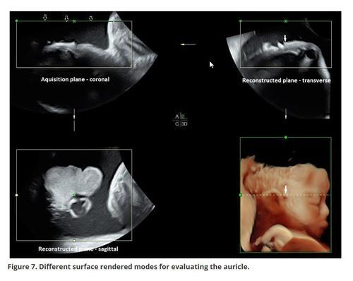
The difficulty with imaging fetal ears is that the information that can be generated on 2D imaging is limited, and 3D images, although not difficult to obtain, requires evaluation of the entire face on a single acquired volume. This is necessary to assess ear location and rotation on the face. The biggest difficulty that I encounter with 3D imaging is the inability to consistently evaluate both ears. The ear in the far field is often difficult or impossible to evaluate and it is well established that both syndromic and non-syndromic ear anomalies are often asymmetrical or unilateral.
Looking ahead, what excites you the most about the future of ultrasound, particularly in your specialized field of fetal ear imaging?
The most exciting and somewhat uncertain future of fetal imaging involves artificial intelligence or AI. AI’s ability to analyze data, access all clinical knowledge in a specific area in real time, as well as its inherent problem-solving techniques has the power to assist in evaluating anomalies. It has the potential to group together a set of unrelated anomalies into a possible syndrome.
The combination of AI and ultrasonography will improve the quality of medical services and patient care, through process efficiencies, scaling and reducing the rates of both misdiagnosis and missed diagnosis.
However, medical ethics, which is critical when conducting any clinical research, may affect the development and application of AI models. Any diagnostic algorithm must consider questions such as is this information correct i.e., peer reviewed, and who bears responsibility and possible medical risk if the information is incorrect.
In order to achieve optimal clinical applications from AI, the diagnostic algorithm must include results of the scan as well as multidimensional information, such as patient age, GA, and medical history. Superior clinical applications and more comprehensive AI models can only be achieved by multidisciplinary collaboration.
AI is not yet capable of considering other social variables that may influence a medical decision.
There are real concerns about what the role of the physician and the sonographer will be as we move towards integrating more AI features into our everyday workstreams.
Read Dr Ian Suchet's free-access VISUOG chapter on Normal ear and its evaluation and discover more about Fetal Ears in a further seven VISUOG chapters contributed by Dr Suchet.