

Ovarian masses: imaging and management is a key topic at this years ISUOG World Congress.
Accurate and timely characterisation of ovarian lesions is essential for guiding appropriate clinical management, with ultrasound assessment remaining central to this process. In an era of increasingly sophisticated imaging and evolving terminology, expert ultrasound evaluation continues to play a pivotal role in differentiating benign from malignant adnexal masses.
This dynamic field continues to evolve, supported by updated definitions and clarifications from the International Ovarian Tumor Analysis (IOTA) group, which aim to standardise the sonographic description of adnexal tumors. Congress sessions will explore the application of pattern recognition in distinguishing benign from malignant masses, the emerging role of artificial intelligence in refining these assessments, and the characteristic ultrasound patterns associated with the principal malignant ovarian tumor groups. International experts in the field will debate how collective imaging features inform standardised risk-prediction models, and which ultrasound markers are associated with non-resectable disease and their implications for preoperative planning.
Amid these developments of advancing evidence and ongoing debate, the 36th ISUOG World Congress in London offers a unique opportunity to engage with cutting-edge research and hear directly from international experts in gynecologic ultrasound imaging and gynecologic oncology. Through scientific sessions and expert-led discussions spanning global perspectives, attendees will deepen their understanding of ovarian lesion assessment, refine their diagnostic approach and explore the controversies shaping contemporary practice.
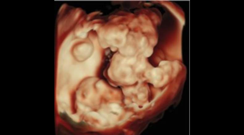
Ovarian mass on transabdominal three-dimensional ultrasound imaging with surface rendering, depicting bizarre-looking central solid component
Dumont, S., Amant, F., Froyman, W., Timmerman, D., Van Rompuy, A.-.-S. and Van den Bosch, T. (2025), Mixed germ cell tumor presenting with mixed sonographic appearance and unique clinical presentation. Ultrasound Obstet Gynecol, 66: 537-540. https://doi.org/10.1002/uog.29179
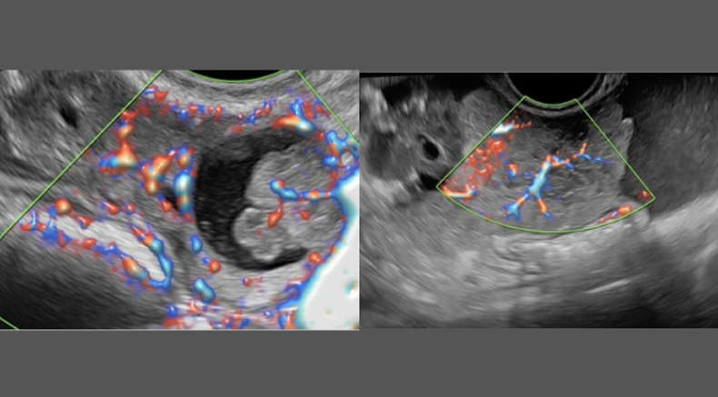
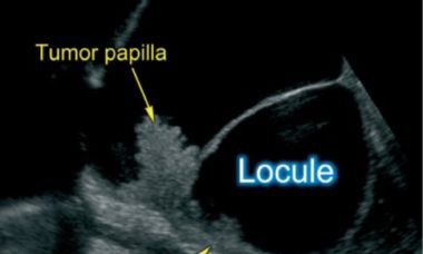
Unilocular-solid cyst with a papillary projection containing microcysts (above). Ovary containing a unilocular-solid cyst with a single papillary projection
Timmerman, D., Valentin, L., Testa, A.C., Froyman, W., Landolfo, C., Kotlarz, A., Fischerova, D., Van Calster, B., Bourne, T. and Collaborators (2026), Terms, definitions and measurements to describe the sonographic features of adnexal tumors: updated consensus opinion
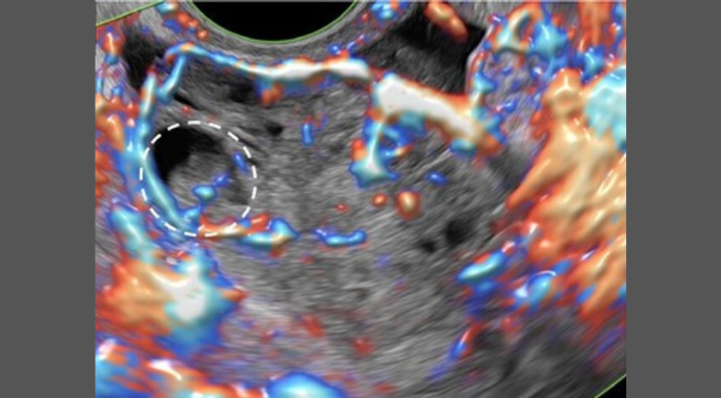
Power Doppler ultrasound image showing a solid tumor with a small cyst containing a papillary projection. Dashed circle surrounds the papillary projection
Timmerman, D., Valentin, L., Testa, A.C., Froyman, W., Landolfo, C., Kotlarz, A., Fischerova, D., Van Calster, B., Bourne, T. and Collaborators (2026), Terms, definitions and measurements to describe the sonographic features of adnexal tumors: updated consensus opinion
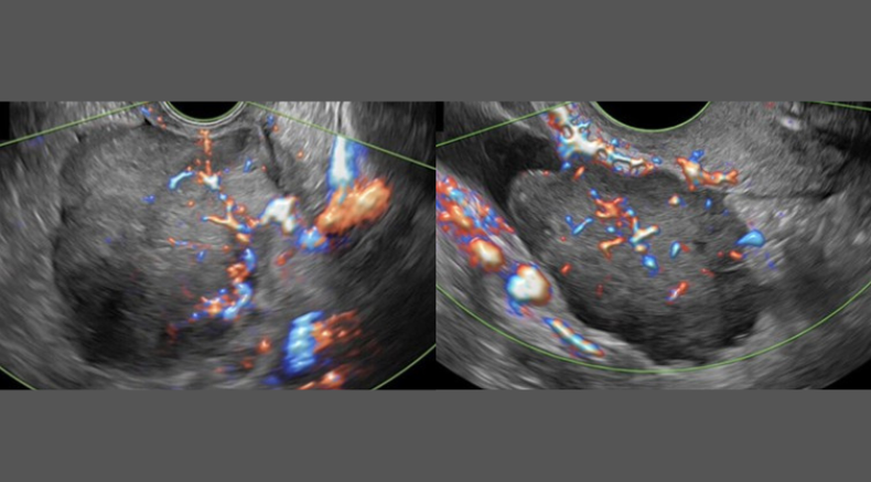
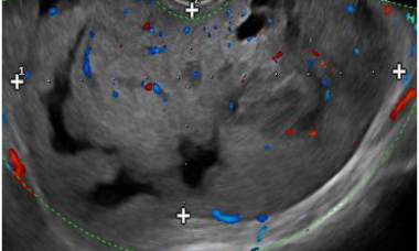
Solid tumor with an irregular gently lobulated surface (above) Solid tumor with spiculated contour(below)
Timmerman, D., Valentin, L., Testa, A.C., Froyman, W., Landolfo, C., Kotlarz, A., Fischerova, D., Van Calster, B., Bourne, T. and Collaborators (2026), Terms, definitions and measurements to describe the sonographic features of adnexal tumors: updated consensus opinion
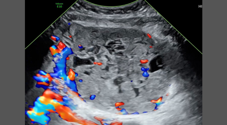
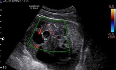
Color Doppler ultrasound images of left-sided multilocular-solid paraovarian tumor
Hovsepyan, L., Malakyan, Z., Saradyan, A., Asilbekyan, N., Mazmanian, I., Eloyan, H. and Valentin, L. (2026), Clinical and sonographic characteristics of adnexal STK11 tumor: newly recognized tumor associated with Peutz–Jeghers syndrome. Ultrasound Obstet Gynecol. https://doi.org/10.1002/uog.70198
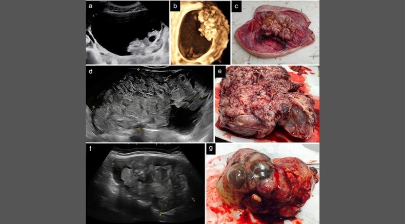
Grayscale ultrasound images (a,d,f), three-dimensional rendering (b) corresponding macroscopic appearance (c,e,g) of three ovarian immature teratomas: (a–c) tumor 1, (d,e) tumor 2 and (f,g) tumor 3
Landolfo, C., Froyman, W., Testa, A.C., Fischerova, D., Franchi, D., Yazbek, J., Saso, S., Fruscio, R., Chiappa, V., Czekierdowski, A., Van Gorp, T., Moro, F., Savelli, L., Zannoni, G.F., Bourne, T., Timmerman, D., Valentin, L. and Collaborators (2026), Imaging in gynecological disease (29)
Why are ovarian masses a key topic at ISUOG 2026?
Sessions where you can learn more about this topic at ISUOG 2026 World Congress
Supplement your learning before Congress
Recent UOG articles
- Imaging in gynecological disease (31): clinical and ultrasound characteristics of ovarian mature cystic teratomas containing malignancies - M. T. Giudice, F. Moro, F. Pozzati, D. Franchi, C. Landolfo, L. Savelli, V. Chiappa, S. Guerriero, G. F. Zannoni, G. Scaglione, W. Froyman, D. Timmerman, T. Bourne, A. Fagotti, L. Valentin, A. C. Testa, Collaborators. First published: 11 May 2026
- Developing and validating ultrasound-based machine-learning models incorporating radiomics features to predict malignancy in adnexal masses
- F. Moro, M. Ciancia, S. di Berardino, G. Baldassari, F. Mascilini, F. Ciccarone, C. Nero, H. E. Tran, T. Pasciuto, L. Boldrini, D. Giannarelli, A. Ledger, T. Bourne, W. Froyman, D. Timmerman, L. Valentin, A. Fagotti, A. C. Testa. First published: 30 March 2026 - The diagnostic puzzle: Meigs' syndrome, pseudo-Meigs' syndrome and ovarian cancer - F. Moro, G. Pellecchia, F. Perrone, N. D'Alessandris, A. Fagotti, A. C. Testa. First published: 06 March 2026
- Clinical and sonographic characteristics of adnexal STK11 tumor: newly recognized tumor associated with Peutz–Jeghers syndrome - L. Hovsepyan, Z. Malakyan, A. Saradyan, N. Asilbekyan, I. Mazmanian, H. Eloyan, L. Valentin. First published: 06 March 2026
- Diagnostic accuracy of ultrasound models for assessment of ovarian tumors: systematic review and meta-analysis - E. Lems, A. H. Koch, E. J. L. G. Delvaux, J. C. Leemans, M. Y. Bongers, C. A. R. Lok, B. L. Ramaekers, P. M. A. J. Geomini. First published: 04 December 2025
- Imaging in gynecological disease (29): clinical and ultrasound features of primary ovarian immature teratoma - C. Landolfo, W. Froyman, A. C. Testa, D. Fischerova, D. Franchi, J. Yazbek, S. Saso, R. Fruscio, V. Chiappa, A. Czekierdowski, T. Van Gorp, F. Moro, L. Savelli, G. F. Zannoni, T. Bourne, D. Timmerman, L. Valentin, Collaborators. First published: 21 October 2025
UOG videos


The diagnostic puzzle: Meigs' syndrome, pseudo-Meigs' syndrome and ovarian cancer

Mixed germ cell tumor presenting with mixed sonographic appearance and unique clinical presentation

Growing teratoma syndrome after treatment of ovarian immature teratoma: ultrasound images of a very rare condition
Lectures

What type of ovarian masses are safe to manage expectantly?
W. Froyman 2025

Using AI in the diagnostic model: the development of ADNEX-AI
D. Timmerman 2025

How confident can you be using ultrasound to stage ovarian malignancy?
D. Fischerova 2025

Ovarian Masses: Challenging cases
J. Preisler Romanow 2025
ISUOG Guideline and Consensus Statements
- Updated Consensus Opinion on the terms, definitions and measurements to describe the sonographic features of adnexal tumors from the International Ovarian Tumor Analysis (IOTA) Group
- ISUOG/ESGO Consensus Statement on ultrasound-guided biopsy in gynecological oncology
- ESGO-ISUOG-IOTA-ESGE Consensus Statement on preoperative diagnosis of ovarian tumors
VISUOG
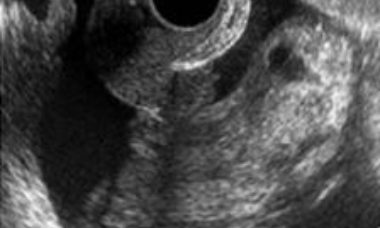
Malignant struma ovarii
Struma ovarii is a rare form of ovarian mature teratoma that contains mostly thyroid tissue. Malignant transformation is uncommon, only about 5% of struma ovarii being malignant. The variable sonographic features of struma ovarii and its rare occurrence makes the sonographic diagnosis very challenging.
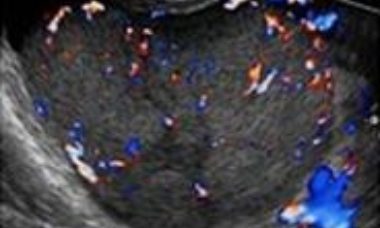
Ovarian dysgerminoma
Dysgerminomas are malignant ovarian germ-cell tumors. Malignant germ-cell tumors of the ovary occur in young women, 75% being diagnosed in the second and third decades of life. At macroscopic evaluation, ovarian dysgerminomas are characteristically solid and well-encapsulated with an average diameter of 15 cm.
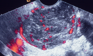
Sertoli-Leydig cell tumors
Sertoli cell tumors, Sertoli-Leydig cell tumors and Leydig cell tumors are sex cord-stromal tumors, and one of the rarest gynecological malignancies, accounting for 0.5-1 % of ovarian tumors. Sertoli cell tumors and Sertoli-Leydig cell tumors are most common in young patients.
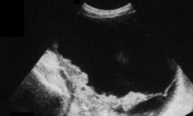
Brenner tumor
Brenner tumors are surface epithelial–stromal tumors of the ovary, which were first described in detail by Fritz Brenner in 1907. Brenner tumors represent 3.2 % of all ovarian tumors. About 99% of them are benign and most patients are postmenopausal. Brenner tumors are usually unilateral.
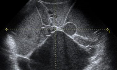
Borderline ovarian tumor (BOT)
Borderline ovarian tumors (BOTs) are epithelial tumors with low grade of malignancy. BOTs account for 10–15% of epithelial ovarian tumors. These tumors occur in younger women, with almost 30% of patients younger than 40 years, and are often diagnosed at an earlier stage than invasive carcinomas.
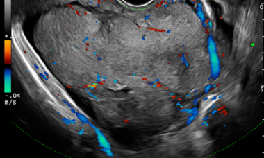
Clear cells carcinoma
Clear cell carcinoma represents 5–25% of all ovarian carcinomas. Tumors can measure up to 30 cm in diameter. In most cases, the cut surface reveals a thick-walled cyst with papillary projections. The histologic patterns include tubulocystic, papillary and solid. The most representative image on ultrasound is a unilateral mass larger than 10 cm with a solid component.
Endometrioid carcinoma
Endometrioid carcinoma represents 10-15% of ovarian epithelial carcinomas. In 15-20% of cases endometrial carcinoma is diagnosed at the same time. Tumors are solid or cystic with a mass protruding into the lumen. The most common microscopic pattern is characterised by a confluent glandular epithelial proliferation.
Mucinous carcinoma
Mucinous carcinomas comprise 2–3% of ovarian carcinomas. Most mucinous carcinomas are well differentiated, containing areas of cystadenoma and atypical proliferative tumor mixed with areas of carcinoma. The size and laterality of the tumor can suggest whether it is primary or metastatic in nature.
Serous carcinoma
Low grade serous carcinoma (LGSC) is a rare disease whereas high grade serous ovarian carcinoma (HGSC) is the most common ovarian malignancy. Non-invasive LGSCs are often bilateral and papillae on the outer surface of the cyst are frequently present. Invasive LGSCs exhibit a papillary growth.
Metastases to the ovary
The ovary is a common site of metastases from malignant tumors. Most metastases in the ovaries originate in the gastrointestinal tract or the breast. The distinction between primary and metastatic ovarian neoplasm is of critical importance, since surgical cytoreduction is the treatment of choice for the former.
Patient information
Ovarian Carcinosarcoma
This leaflet is to help you understand what an ovarian carcinosarcoma is, what tests you need and the implication of being diagnosed are for you, your baby and your family.
Metatasis of the Ovary
This leaflet is to help you understand what Metastases of the Ovary is, what tests you need and the implication of being diagnosed with Metastases of the Ovary for you, your baby and your family.
Metastatic Lesions
This leaflet is to help you understand what Metastatic Lesions are, what causes them, what tests you need and what the implications of being diagnosed with a tumor.
Malignant struma ovarii
This leaflet is to help you understand what Malignant struma ovarii is, how does it happen, what tests you need and what are the long term implications of the diagnosis.
Granulosa cell tumor
This leaflet is to help you understand what Granulosa cell tumor is, how does it happen, what tests you need and what are the long term implications of the diagnosis.
Epithelial ovarian carcinoma
This leaflet is to help you understand what Epithelial ovarian carcinoma is, what tests you need and the implication of being diagnosed with Epithelial ovarian carcinoma for you, your baby and your family.
Borderline Ovarian Tumor
This leaflet is to help you understand what Borderline Ovarian Tumor is, how does it happen, what tests you need and what are the long term implications of the diagnosis.
CME activities
- CME Activity: Simple Ultrasound-based Rules for the Diagnosis of Ovarian Cancer
- CME Activity: Ovarian Cancer Staging
- CME Activity - Managing Ovarian Masses
- CME Activity: Masterclass: Evaluating Metastatic Disease in Ovarian Cancer and Tumor Extension in Endometrial and Cervical Disease
Key questions
Which sonographic features are most useful in distinguishing benign from malignant adnexal masses?
What do the updated IOTA definitions add to the standardised description and reporting of adnexal tumors?
Which ultrasound markers suggest non-resectable disease, and how should these findings inform preoperative planning?
How might emerging artificial intelligence tools refine adnexal mass assessment and improve diagnostic accuracy in gynecologic ultrasound?