Supplement your learning for ISUOG's education course on exploring the fetal brain.
Join course Director Prof. Francesco D'Antonio, and co-Chairs Prof. Asma Khalil and Prof. Simon Meagher for this engaging and informative course. Together with assistance from an expert panel of speakers they will dive into exploring the fetal brain taking you through the imaging, prognosis and counselling process. This course offers attendees a valuable opportunity to gain a comprehensive understanding of the fetal brain and how to approach complex practical situations.
Learning Objectives:
- Gain expertise in basic and advance assessment of fetal brain
- Improve your knowledge of central nervous system anomalies
- Become familiar with the imaging techniques of fetal brain assessment (ultrasound and MRI)
- Engage with an international panel of experts on topics such as peculiar posterior fossa, cortical anomalies, infections
- View complex clinical cases in fetal neurology
- Hear more about how to counsel women with fetal brain anomalies
- Discover new approaches to identify foetuses at risk of adverse neurodevelopmental outcome
- Learn how to identify subtle central nervous system anomalies
Explore the topic before you attend our course
In order to make the most of this learning experience and help you achieve your learning objectives, we have prepared a path to guide you from the essentials to our course’s topics through ISUOG resources. The material below, will take you from the most basics to a more comprehensive view of the fetal brain through imaging, prognosis and counselling, some open to everyone and some available only to ISUOG members –some may even grant you CME points:
Some of these activities are exclusively available to our members. Become a member today.
VISUOG
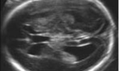
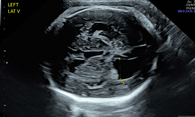
Mild Ventriculomegaly
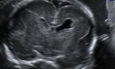
There is marked biologic variability in the size of the fetal lateral ventricles. Empiric data suggests that fetuses with a width of the atrium (or posterior horn) of lateral ventricles of 10 mm or more have an increased risk of abnormal outcome. When the measurement is 10-15mm the risk is relatively low.
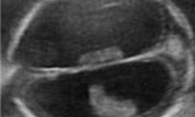
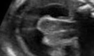
Severe Ventriculomegaly
The current definition is an increased size of the lateral ventricles, with a transverse diameter of the atrium in excess of 15mm without evidence of other cerebral malformations. It is a rare condition, usually a part of complex cerebral abnormalities, less frequently the consequence of obstructed cerebrospinal fluid turnover.
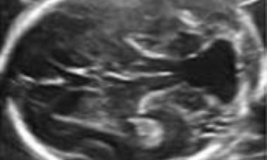
Intracranial Hemorrage
Intracranial hemorrage may occur within the lateral ventricles or in the subdural space. The sonographic appearance changes with time. An echogenic collection is first seen, and in the following days it develops into a complex mass frequently complicated by sever ventriculomegaly.
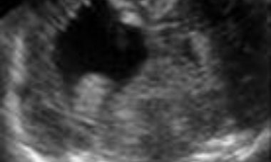
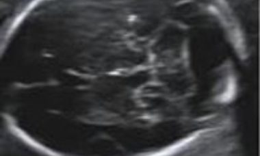
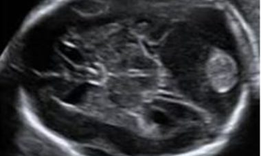
Porencephaly
Porencephaly is characterized by single or multiple cysts replacing the brain parenchyma. The cyst may communicate with the lateral ventricle, subarachnoid space or both. It is usually a sporadic condition caused by hemorrage, ischemia or infections.
Aqueductal Stenosis
Aqueductal stenosis (AS) results from a narrowing or occlusion of the aqueduct of Sylvius.
Holoprosencephaly
Holoprosencephaly derives from failure of separation of the cerebral hemispheres. In the most severe forms there is one undivided cerebral mass that contains a crescent shaped rudimentary ventricular cavity. In these cases, severe cranio-facial anomalies (cyclopia, hypotelorism, median cleft face) are associated.
Dandy-Walker Complex
Under this term are included a group of conditions that share in common one sonographic findings: the impression that the fourth ventricle communicates with the cisterna magna. These conditions include: Dandy-Walker malformation, Blake’s pouch cyst, vermian hypoplasia/agenesis. They have a similar sonographic appearance, particularly in early gestation, and differentiation requires a multiplanar approach.
Megacisterna Magna
Enlarged (> 10 mm) cisterma magna with a normal cerebellum. It may be associated with other anomalies, including trisomy 18 and other cerebral malformations. When isolated, fetal mega cisterna magna has a good chance of intrauterine remission, and is usually (> 90%of cases) associated with a good outcome.
Disorders of neuronal migration: Lissencephaly, Cobblestone Malformations and Heterotopia
Malformations of cortical development (MCDs) are neurodevelopmental disorders that result from abnormal development of the cerebral cortex in utero secondary to genetic, infectious or vascular causes.
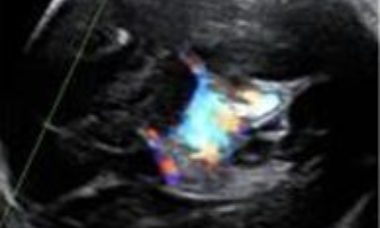
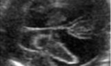
Enlargement of the vein of Galen
It is the consequence of two types of arteriovenous malformation: the vein of Galen aneurysmal malformation and the pial arteriovenous malformation. The sonographic appearance is similar and the most prominent finding is the enlargement of the vein of Galen, that on Color Doppler contains turbulent blood flow.
Dural Sinuses Thrombosis
Thrombosis of the dural sinus occurs rarely in intrauterine life. It is characterized by the dilatation of the most inferior and posterior portion of the superior sagittal sinus and of the sinus confluence, that appear as a fluid collection in the occipital area of the fetal cranium.
UOG Articles
Fetal corpus callosum anomalies: from disease of classification to classification of disease
L. J. Salomon and D. Paladini
‘Choroid bar’: easy-to-seek marker of normal posterior fossa at 12–14 weeks’ gestation
D. Paladini, G. Biancotto, F.Della Sala and P. V. Acharya
Patient Information
Septo-optic Dysplasia (SOD)
This leaflet is to help you understand what septo-optic dysplasia (SOD) is, what causes it, and the implication of being diagnosed for your baby after it is born.
Cytomegalovirus (CMV) infection in pregnancy
This leaflet is to help you understand what Cytomegalovirus (CMV) infection is, what tests you need and the implication of having been diagnosed with Cytomegalovirus (CMV) infection for you, your baby and your family.
Cerebellar hypoplasia
This leaflet is to help you understand what Cerebellar hypoplasia is, what tests you need, and the implication of having been diagnosed with Cerebellar hypoplasia for you, your baby and your family.
Rhombencephalosynapsis (RES)
This leaflet is to help you understand what Rhombencephalosynapsis (RES) is, what tests you need, and the implication of being diagnosed for you and your baby.