Supplement your learning for the ISUOG Course 'ISUOG advanced neurosonography course: the dark side of the brain and changing the paradigm' with the following learning resources.
ISUOG is pleased to announce our next education course on neurosonography. It will be taking place on Saturday 21 August 2021 via livestream.
Explore the topic before you attend our course:
In order to make the most of this learning experience and help you achieve your learning objectives, we have prepared a path to guide you from the essentials to our course’s topics through ISUOG resources. The material below will take you from the most basic aspects to a more comprehensive view of the course material, and some activities may grant you CME points.
Some of these activities are exclusively available to our members. Become a member today.
ISUOG Guidelines
ISUOG Practice Guidelines (updated): sonographic examination of the fetal central nervous system. Part 1: performance of screening examination and indications for targeted neurosonography
ISUOG Practice Guidelines (updated): sonographic examination of the fetal central nervous system. Part 2: performance of targeted neurosonography
Performance of fetal magnetic resonance imaging
CME Activity - ISUOG Practice Guidelines: Performance of Fetal Magnetic Resonance Imaging
VISUOG
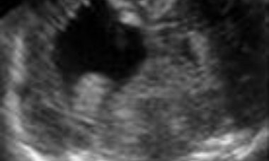
Holoprosencephaly
Holoprosencephaly derives from failure of separation of the cerebral hemispheres. In the most severe forms there is one undivided cerebral mass that contains a crescent shaped rudimentary ventricular cavity. In these cases, severe cranio-facial anomalies (cyclopia, hypotelorism, median cleft face) are associated.
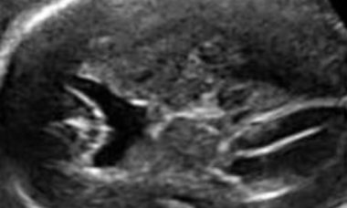
Agenesis of Septum Pellucidum
Absence of the septum pellucidum is found with many cerebral malformations, including holoprosencephaly, agenesis of corpus callosum, ventriculomegaly, open spina bifida, cortical malformations. It may be an isolated abnormality and in this case the cerebral anatomy is unremarkable but for fusion of the frontal horns.
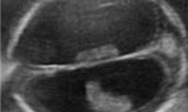
Severe Ventriculomegaly
The current definition is an increased size of the lateral ventricles, with a transverse diameter of the atrium in excess of 15mm without evidence of other cerebral malformations. It is a rare condition, usually a part of complex cerebral abnormalities, less frequently the consequence of obstructed cerebrospinal fluid turnover.
Intracranial Hemorrage
Intracranial hemorrage may occur within the lateral ventricles or in the subdural space. The sonographic appearance changes with time. An echogenic collection is first seen, and in the following days it develops into a complex mass frequently complicated by sever ventriculomegaly. In the most severe forms the hemoragge may be complicated by an infarct in the brain parenchyma. The etiology is variable.
Porencephaly
Porencephaly is characterized by single or multiple cysts replacing the brain parenchyma. The cyst may communicate with the lateral ventricle, subarachnoid space or both. It is usually a sporadic condition caused by hemorrage, ischemia or infections.
Dandy-Walker Complex
Under this term are included a group of conditions that share in common one sonographic findings: the impression that the fourth ventricle communicates with the cisterna magna. These conditions include: Dandy-Walker malformation, Blake’s pouch cyst, vermian hypoplasia/agenesis. They have a similar sonographic appearance, particularly in early gestation, and differentiation requires a multiplanar approach.
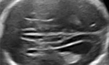
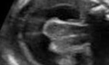
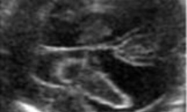
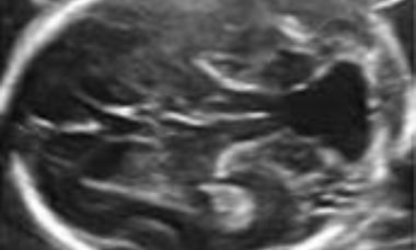
Agenesis of the Corpus Callosum
Agenesis of the corpus callosum may be either complete or partial. Intracranial anatomy is variable. Other intracranial anomalies are frequently encountered including ventriculomegaly, cysts, lipomas. It may be a part of genetic and genetic syndromes. The prognosis is uncertain.
UOG articles
Supplement your learning with specially chosen articles from UOG.
P. Volpe, R. De Robertis, T. Fanelli, S. Boito, G. Volpe, C. Votino, N. Persico, R. Chaoui
14 June 2021
H. Mahallati, A. Sotiriadis, C. Celestin, A. E. Millischer, P. Sonigo, D. Grevent, N. O'Gorman, N. Bahi-Buisson, T. Attié-Bitach, Y. Ville, L. J. Salomon
15 August 2020
D Paladini
26 June 2021
N. Volpe, A. Dall'Asta, E. Di Pasquo, T. Frusca, T. Ghi
13 October 2020
CME Activity: Neurodevelopmental Disorder in Children Believed to Have Isolated Mild Ventriculomegaly Prenatally
CME Activity: Outcome of Fetuses with Prenatal Diagnosis of Isolated Severe Bilateral Ventriculomegaly: Systematic Review and Meta-Analysis
Learning Modules
R Pooh 2021
D Paladini 2021
J Copel 2021
S Meagher 2017
CME Activity: Fetal Brain Tumors & Cysts
CME Activity: What is Wrong with this Cavum Septi Pellucidi?
CME Activity: Facts and Myths about the Cerebellar Vermis & Cisterna Magna
CME Activity - Fetal Neurosonography: Tips and Tricks