Supplement your learning on Ultrasound in uterine malformations and pathology
Join us for this engaging and informative course. Together with a panel of expert speakers from around the world, they will dive into the latest discourse surrounding Ultrasound in uterine malformations and pathology. This course offers attendees a valuable opportunity to gain a comprehensive understanding of the new advances in this area and learn how to apply them in practical situations.
Learning Objectives Malformations:
- Understand embryology and classification of uterine malformations.
- Identify uterine anomalies using 2D and 3D ultrasound.
- Differentiate between different types of uterine anomalies.
- Correlate ultrasound findings with MRI and clinical implications.
- To learn apply knowledge to infertility, obstetric outcomes, and management decisions.
Learning Objectives Pathology:
- Develop expertise in the ultrasound evaluation of endometrial pathology
- Gain expertise in ultrasound diagnosis of myometrial pathology
- Learn to differentiate benign from malignant uterine findings
- Enhance understanding of the management of symptomatic and asymptomatic women with sonographic findings in the uterus
- Gain skills in addressing diagnostic and management challenges in uterine pathology
- Expand knowledge of the roles of fibroids, adenomyosis, and endometrial pathology in infertility
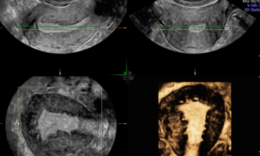
- Understand the complementary roles of ultrasound and MRI in diagnosing adenomyosis
Explore the topic before you attend our course
In order to make the most of this learning experience and help you achieve your learning objectives, we have prepared a path to guide you from the essentials to our course’s topics through ISUOG resources. The material below, will take you from the most basics to a more comprehensive view of Fetal Growth Restriction: From AI-Powered Prediction to Precision Medicine, some open to everyone and some available only to ISUOG members –some may even grant you CME points:
Some of these activities are exclusively available to our members. Become a member today.
ISUOG Guideline
T. Van den Bosch, R. Heremans, C. Landolfo, E. Epstein, F. P. G. Leone, T. Bourne, D. Timmerman, Collaborators
First published: 16 January 2026
VISUOG
Hemi-uterus
The development of a hemi-uterus, also called unicornuate uterus, is due to complete or partial agenesis of a unilateral Mullerian duct.
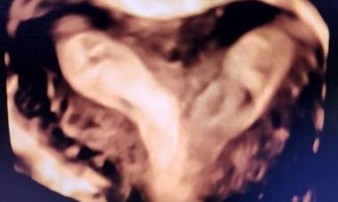
Septate Uterus
Septate uterus is a congenital uterine anomaly due to defective resorption of the medial fusion of the Müllerian ducts. The uterus has a normal external uterine morphology and a septum dividing the uterine cavity. It is further classified as partial or complete septate uterus.
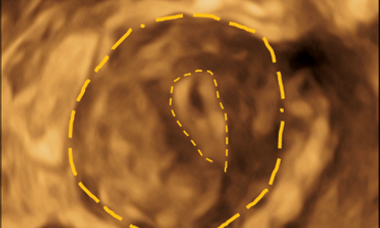
Bicorporeal uterus
Bicorporeal uterus is characterized by the presence of an external indentation at the fundal midline that is greater than 50% of the thickness of the uterine wall.
Dysmorphic Uterus
A dysmorphic uterus includes subtypes T-shaped uterus and infantile uterus. It can be acquired or congenitally present.
Uterine fibroids
Uterine fibroids, also called myomas or leiomyomas, are benign fibromuscular tumours of the myometrium.
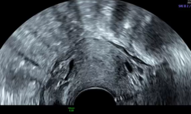
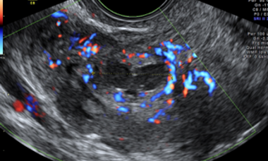
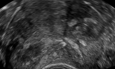
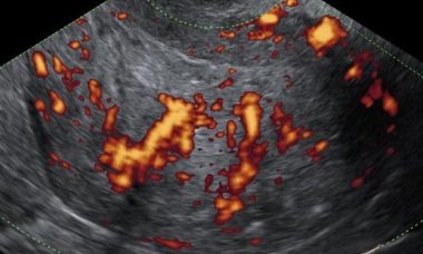
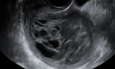
Adenomyosis
Sonographic features considered typical for adenomyosis are echogenic subendometrial lines and buds, hyperechogenic islands, myometrial cysts, fan‐shaped shadowing, an irregular or interrupted junctional zone, translesional vascularity, asymmetrical thickening of the myometrium, and/or an enlarged globular uterus.
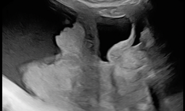
Intrauterine adhesions
Intrauterine adhesions (IUA) are defined as abnormal fibrous bands bridging opposing sides of the endometrial cavity, causing obliteration and obstruction of the cavity and/or cervical canal.
Endometrial Cancer
Endometrial cancer is the most common malignancy of the female genital tract, affecting 2-3% of women during their lifetime. The median age at diagnosis is 60-65 years and the most commonly associated risk-factor is obesity.
Uterine sarcomas
Uterine sarcomas are rare malignant tumors arising from the mesenchymal tissues of the uterus, i.e. the endometrial stroma, uterine muscle and connective tissue. They represent 1% of female genital tract malignancies and 3-7% of all uterine malignances.
Uterine and endometrial masses from other primary tumors
Extragenital primary tumors that metastasize to the endometrium and or uterine cervix are infrequent conditions and represent a diagnostic challenge among clinicians and pathologists.
Patient Information
Septate Uterus
This leaflet is to help you understand what Septate Uterus is, what tests you need, and the implication of being diagnosed for you.
Hemi-uterus
This leaflet is to help you understand what Hemi-uterus is, how does it happen, what tests you need and what are the long term implications of the diagnosis.
Bicorporeal uterus
This leaflet is to help you understand what Bicorporeal uterus is, how does it happen, what tests you need and what are the long term implications of the diagnosis.
Uterine Fibroids
This leaflet is to help you understand what Uterine Fibroids are, the associated symptoms, and what treatments are available to you.
Adenomyosis
This leaflet is to help you understand what adenomyosis is, what tests you need, and the implication of being diagnosed for you and your baby.
Intrauterine Adhesions
This leaflet is to help you understand what intrauterine Adhesion is, what tests you need, and the implication of being diagnosed.
Endometrial hyperplasia
This leaflet is to help you understand what endometrial hyperplasia is, what tests you need and the implication of being diagnosed, and how it can be treated.
Endometrial Cancer
This leaflet is to help you understand what endometrial cancer is and what causes it, how it can be diagnosed and the treatments available to you.
UOG Articles
S. Nijjar, S. Kastora, A. Bajrami, S. Solangon, M. Widschwendter, D. Jurkovic
First published: 04 November 2025
Ultrasound features using MUSA terms and definitions in uterine sarcoma and leiomyoma: cohort study
C. De Bruyn, J. Ceusters, K. Vanden Brande, S. Timmerman, W. Froyman, D. Timmerman, A.-S. Van Rompuy, A. Coosemans, T. Van den Bosch
First published: 16 November 2023
ASRM Müllerian Anomalies Classification 2021: a critical review
A. Ludwin, S. Tudorache, W. P. Martins
First published: 09 June 2022
M. J. Harmsen, T. Van den Bosch, R. A. de Leeuw, M. Dueholm, C. Exacoustos, L. Valentin, W. J. K. Hehenkamp, F. Groenman, C. De Bruyn, C. Rasmussen, L. Lazzeri, L. Jokubkiene, D. Jurkovic, J. Naftalin, T. Tellum, T. Bourne, D. Timmerman, J. A. F. Huirne
First published: 29 September 2021
Definition, prevalence, clinical relevance and treatment of T-shaped uterus: systematic review
M. A. Coelho Neto, A. Ludwin, F. Petraglia, W. P. Martins
First published: 08 September 2020
A. Ludwin, I. Ludwin, M. A. Coelho Neto, C. O. Nastri, B. Bhagavath, S. R. Lindheim, W. P. Martins
First published: 11 April 2019
Imaging in gynecological disease (15): clinical and ultrasound characteristics of uterine sarcoma
M. Ludovisi, F. Moro, T. Pasciuto, S. Di Noi, S. Giunchi, L. Savelli, M. A. Pascual, P. Sladkevicius, J. L. Alcazar, D. Franchi, R. Mancari, M. C. Moruzzi, D. Jurkovic, V. Chiappa, S. Guerriero, C. Exacoustos, E. Epstein, F. Frühauf, D. Fischerova, R. Fruscio, F. Ciccarone, G. F. Zannoni, G. Scambia, L. Valentin, A. C. Testa
First published: 25 March 2019
Videos
Diagnosis of uterine malformations
P. Bortoletto 2025
Differentiating fiboids from sarcomas
A. Testa 2023
What we have learnt from the IETA trial: the appearances associated with different kinds of endometrial pathology
T. Van den Bosch 2023
Diagnosing adenomyosis: what is its clinical relevance and how to treat it
T. Tellum 2023
Gynecological problems associated with Caesarean section scars: when and how do you intervene?
J. Huirne 2023