Supplement your learning for Assessment of adnexal masses using ultrasound.
Join us for this engaging and informative course. Together with a panel of expert speakers from around the world, they will dive into the latest discourse surrounding ultrasound from the First to Third Trimester. This course offers attendees a valuable opportunity to gain a comprehensive understanding of the new advances in this area and learn how to apply them in practical situations.
Learning Objectives:
- To learn simple methods for assessing adnexal masses on ultrasound
- To gain knowledge on how to use the ADNEX model in clinical practice
- To learn how to use O-RADS in patient management
- To improve knowledge on the use of pattern recognition when assessing adnexal masses
- To gain knowledge on the role of ultrasound in diagnosing invasive ovarian malignancies, metastases in the ovaries and rare ovarian tumours
- To gain knowledge on the role of ultrasound in identifying non-gynecological pathology
Explore the topic before you attend our course
In order to make the most of this learning experience and help you achieve your learning objectives, we have prepared a path to guide you from the essentials to our course’s topics through ISUOG resources. The material below, will take you from the most basics to a more comprehensive view of Ultrasound meets genetics, some open to everyone and some available only to ISUOG members –some may even grant you CME points:
Some of these activities are exclusively available to our members. Become a member today.
VISUOG
Malignant struma ovarii
Struma ovarii is a rare form of ovarian mature teratoma that contains mostly thyroid tissue. Malignant transformation is uncommon, only about 5% of struma ovarii being malignant. The variable sonographic features of struma ovarii and its rare occurrence makes the sonographic diagnosis very challenging.

Ovarian dysgerminoma
Dysgerminomas are malignant ovarian germ-cell tumors. Malignant germ-cell tumors of the ovary occur in young women, 75% being diagnosed in the second and third decades of life. At macroscopic evaluation, ovarian dysgerminomas are characteristically solid and well-encapsulated with an average diameter of 15 cm.
Sertoli-Leydig cell tumors
Sertoli cell tumors, Sertoli-Leydig cell tumors and Leydig cell tumors are sex cord-stromal tumors, and one of the rarest gynecological malignancies, accounting for 0.5-1 % of ovarian tumors. Sertoli cell tumors and Sertoli-Leydig cell tumors are most common in young patients.
Brenner tumor
Brenner tumors are surface epithelial–stromal tumors of the ovary, which were first described in detail by Fritz Brenner in 1907. Brenner tumors represent 3.2 % of all ovarian tumors. About 99% of them are benign and most patients are postmenopausal. Brenner tumors are usually unilateral.
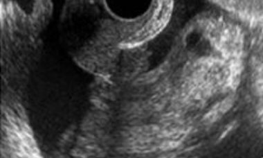
Borderline ovarian tumor (BOT)
Borderline ovarian tumors (BOTs) are epithelial tumors with low grade of malignancy. BOTs account for 10–15% of epithelial ovarian tumors. These tumors occur in younger women, with almost 30% of patients younger than 40 years, and are often diagnosed at an earlier stage than invasive carcinomas.
Clear cells carcinoma
Clear cell carcinoma represents 5–25% of all ovarian carcinomas. Tumors can measure up to 30 cm in diameter. In most cases, the cut surface reveals a thick-walled cyst with papillary projections. The histologic patterns include tubulocystic, papillary and solid. The most representative image on ultrasound is a unilateral mass larger than 10 cm with a solid component.
Endometrioid carcinoma
Endometrioid carcinoma represents 10-15% of ovarian epithelial carcinomas. In 15-20% of cases endometrial carcinoma is diagnosed at the same time. Tumors are solid or cystic with a mass protruding into the lumen. The most common microscopic pattern is characterised by a confluent glandular epithelial proliferation.
Mucinous carcinoma
Mucinous carcinomas comprise 2–3% of ovarian carcinomas. Most mucinous carcinomas are well differentiated, containing areas of cystadenoma and atypical proliferative tumor mixed with areas of carcinoma. The size and laterality of the tumor can suggest whether it is primary or metastatic in nature.
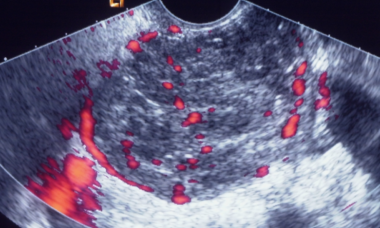
Serous carcinoma
Low grade serous carcinoma (LGSC) is a rare disease whereas high grade serous ovarian carcinoma (HGSC) is the most common ovarian malignancy. Non-invasive LGSCs are often bilateral and papillae on the outer surface of the cyst are frequently present. Invasive LGSCs exhibit a papillary growth.
Metastases to the ovary
The ovary is a common site of metastases from malignant tumors. Most metastases in the ovaries originate in the gastrointestinal tract or the breast. The distinction between primary and metastatic ovarian neoplasm is of critical importance, since surgical cytoreduction is the treatment of choice for the former.
Extrapelvic sites of Endometriosis
Extrapelvic endometriosis most often affects the gastrointestinal tract, umbilicus, inguinal area, cesarean scar, diaphragm and pelvic nerves. The diagnosis is challenging and imaging methods can be used to access suspected lesions, and to evaluate the pelvic cavity since isolated extraperitoneal endometriosis is rare.
Deep Endometriosis
Explore chapters on deep endometriosis
Patient Information
Borderline Ovarian Tumor
This leaflet is to help you understand what Borderline Ovarian Tumor is, how does it happen, what tests you need and what are the long term implications of the diagnosis.
Epithelial ovarian carcinoma
This leaflet is to help you understand what Epithelial ovarian carcinoma is, what tests you need and the implication of being diagnosed with Epithelial ovarian carcinoma for you, your baby and your family.
Granulosa cell tumor
This leaflet is to help you understand what Granulosa cell tumor is, how does it happen, what tests you need and what are the long term implications of the diagnosis.
Malignant struma ovarii
This leaflet is to help you understand what Malignant struma ovarii is, how does it happen, what tests you need and what are the long term implications of the diagnosis.
Metastatic Lesions
This leaflet is to help you understand what Metastatic Lesions are, what causes them, what tests you need and what the implications of being diagnosed with a tumor.
Metatasis of the Ovary
This leaflet is to help you understand what Metastases of the Ovary is, what tests you need and the implication of being diagnosed with Metastases of the Ovary for you, your baby and your family.
Ovarian Carcinosarcoma
This leaflet is to help you understand what an ovarian carcinosarcoma is, what tests you need and the implication of being diagnosed are for you, your baby and your family.
Endometriomas
This leaflet is to help you understand what Endometriosis is, how does it happen, what tests you need and what are the long term implications of the diagnosis?
Anterior compartment endometriosis
This leaflet is to help you understand what anterior compartment endometriosis is, what tests you need and the implication of being diagnosed, as well as the treatment options available to you.
Extra-pelvic endometriosis sites
This leaflet is to help you understand what Endometriosis is, how does it happen, what tests you need and what are the long term implications of the diagnosis?
UOG Articles
F. Pozzati, F. Moro, J. L. Alcazar, K. De Votch, E. Fontana, A. C. Testa
June 2025
F. Mascilini, F. Moro, T. Pasciuto, P. Sladkevicius, W. Froyman, L. Jokubkiene, C. Van Holsbeke, D. Franchi, E. Epstein, S. Guerriero, V. Chiappa, F. Buonomo, M. J. Kudla, J. L. Alcázar, L. Hochberg, F. Ciccarone, L. Quagliozzi, G. Scambia, D. Timmerman, L. Valentin, A. C. Testa
May 2025
Mixed germ cell tumor presenting with mixed sonographic appearance and unique clinical presentation
S. Dumont, F. Amant, W. Froyman, D. Timmerman, A.-S. Van Rompuy, T. Van den Bosch
January 20205
F. Moro, M. Vagni, H. E. Tran, F. Bernardini, F. Mascilini, F. Ciccarone, C. Nero, D. Giannarelli, L. Boldrini, A. Fagotti, G. Scambia, L. Valentin, A. C. Testa
May 2024
F. Moro, M. T. Giudice, G. Bolomini, M. C. Moruzzi, F. Mascilini, L. Quagliozzi, F. Ciccarone, G. Scambia, A. Fagotti, L. Valentin, A. C. Testa
September 2023
R. Heremans, L. Valentin, P. Sladkevicius, S. Timmerman, F. Moro, C. Van Holsbeke, E. Epstein, A. C. Testa, D. Timmerman, W. Froyman
March 2022
UOG Video Clips
Retroperitoneal cyst with iliac stent involvement as primary manifestation of cystic echinococcosis
Growing teratoma syndrome after treatment of ovarian immature teratoma: ultrasound images of a very rare condition
Serous surface papillary borderline ovarian tumors
Diagnostic accuracy of ultrasound signs for detecting adnexal torsion: systematic review and meta-analysis
Learning Modules
Lessons learned from the imaging in gynecology series
A.Testa 2024
Relevance of ovarian cysts in pregnancy
T. Bourne 2024
Ovarian Masses and AI
A.Testa 2023
Machine Learning and Radiomics to classify ovarian pathology
J.Barcroft 2023