
Supplement your learning on Ultrasound excellence in Gynecological malignancies
Join us for this engaging and informative course. Together with a panel of expert speakers from around the world, they will dive into the most challenging and evolving aspects of Ultrasound excellence in Gynecological malignancies. This course offers attendees a valuable opportunity to gain a comprehensive understanding of the new advances in this area and learn how to apply them in practical situations.
Learning objectives:
- Develop a comprehensive understanding of pelvic anatomy and the sonographic features of normal and malignant gynaecological tissues.
- Apply ultrasound techniques to evaluate suspected ovarian, endometrial, cervical, vulvar, and vaginal malignancies, including tumour characterisation and assessment of disease extent.
- Identify sonographic features associated with benign and malignant adnexal masses, and interpret findings using established classification systems and risk models.
- Demonstrate knowledge of the role of ultrasound in staging, treatment planning, and guiding invasive procedures in gynaecological oncology.
- Integrate evidence-based ultrasound approaches into clinical practice to enhance diagnostic accuracy, support multidisciplinary decision-making, and optimise patient care.
Explore the topic before you attend our course
In order to make the most of this learning experience and help you achieve your learning objectives, we have prepared a path to guide you from the essentials to our course’s topics through ISUOG resources. The material below, will take you from the most basics to a more comprehensive view of Ultrasound excellence in Gynecological malignancies, some open to everyone and some available only to ISUOG members –some may even grant you CME points:
Some of these activities are exclusively available to our members. Become a member today.
VISUOG
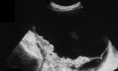
Epithelial origin
Explore the sub-sections on Invasive epithelial tumors, Benign epithelial tumors and Transitional cells tumors.
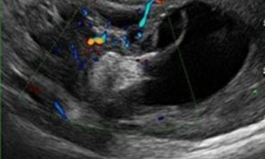
Germ cells tumors
Explore the sub-sections on Rare germ cells tumors, Benign germ cells tumors and Malignant germ cells.
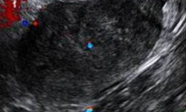
Stromal and sex-cord cells tumors
Explore a sub-section on specialized stromal cell tumors and a chapter on ovarian fibroma and fibrothecoma.
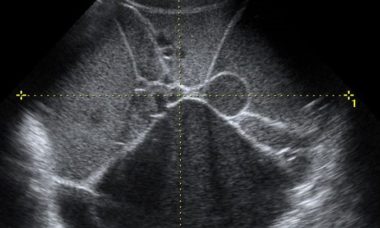
Borderline ovarian tumor (BOT)
Borderline ovarian tumors (BOTs) are epithelial tumors with low grade of malignancy. BOTs account for 10–15% of epithelial ovarian tumors. These tumors occur in younger women, with almost 30% of patients younger than 40 years, and are often diagnosed at an earlier stage than invasive carcinomas.
Metastases to the ovary
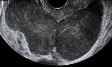
The ovary is a common site of metastases from malignant tumors. Most metastases in the ovaries originate in the gastrointestinal tract or the breast. The distinction between primary and metastatic ovarian neoplasm is of critical importance, since surgical cytoreduction is the treatment of choice for the former.
Endometrial Cancer
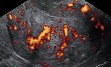
Endometrial cancer is the most common malignancy of the female genital tract, affecting 2-3% of women during their lifetime. The median age at diagnosis is 60-65 years and the most commonly associated risk-factor is obesity.
Uterine sarcomas
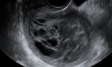
Uterine sarcomas are rare malignant tumors arising from the mesenchymal tissues of the uterus, i.e. the endometrial stroma, uterine muscle and connective tissue. They represent 1% of female genital tract malignancies and 3-7% of all uterine malignances.
ISUOG guidelines and Consensus statements
Patient Information
Borderline Ovarian Tumor
This leaflet is to help you understand what Borderline Ovarian Tumor is, how does it happen, what tests you need and what are the long term implications of the diagnosis.
Epithelial ovarian carcinoma
This leaflet is to help you understand what Epithelial ovarian carcinoma is, what tests you need and the implication of being diagnosed with Epithelial ovarian carcinoma for you, your baby and your family.
Granulosa cell tumor
This leaflet is to help you understand what Granulosa cell tumor is, how does it happen, what tests you need and what are the long term implications of the diagnosis.
Metastatic Lesions
This leaflet is to help you understand what Metastatic Lesions are, what causes them, what tests you need and what the implications of being diagnosed with a tumor.
Malignant struma ovarii
This leaflet is to help you understand what Malignant struma ovarii is, how does it happen, what tests you need and what are the long term implications of the diagnosis.
Cervical cancer
This leaflet is to help you understand what cervical cancer is, what tests you need and the implication of being diagnosed and what treatment options are available to you.
Metatasis of the Ovary
This leaflet is to help you understand what Metastases of the Ovary is, what tests you need and the implication of being diagnosed with Metastases of the Ovary for you, your baby and your family.
Endometrial Cancer
This leaflet is to help you understand what endometrial cancer is and what causes it, how it can be diagnosed and the treatments available to you.
UOG Articles
|
Decidualized ovarian endometrioma mimicking malignancy in a non-pregnant woman S. Alson, S. Neringer, L. Valentin First published: 22 June 2026
M. T. Giudice, F. Moro, F. Pozzati, D. Franchi, C. Landolfo, L. Savelli, V. Chiappa, S. Guerriero, G. F. Zannoni, G. Scaglione, W. Froyman, D. Timmerman, T. Bourne, A. Fagotti, L. Valentin, A. C. Testa, Collaborators First published: 11 May 2026
F. Moro, M. Ciancia, S. di Berardino, G. Baldassari, F. Mascilini, F. Ciccarone, C. Nero, H. E. Tran, T. Pasciuto, L. Boldrini, D. Giannarelli, A. Ledger, T. Bourne, W. Froyman, D. Timmerman, L. Valentin, A. Fagotti, A. C. Testa First published: 30 March 2026
F. Ciccarone, A. Rizzi, A. Biscione, G. Baldassari, E. H. Tran, T. Pasciuto, F. Moro, G. Zinicola, F. Buonomo, M. Colombin, F. Ghezzi, J. Casarin, R. Mancari, F. Borella, A. Kardhashi, M. Roccio, L. Savelli, R. Cioffi, F. Fanfani, G. Ferrandina, G. Scambia, L. Valentin, D. Lorusso, A. C. Testa First published: 06 March 2026
E. Trimarchi, C. Caia, C. Borso, L. Galli, S. Palladino, L. Zanchi, B. Paolini, V. Chiappa First published: 07 September 2025
D. Fischerova, E. Gatti, C. Culcasi, Z. Ng, G. Szabó, L. Zanchi, A. Burgetova, O. Nanka, G. Gambino, M. R. Kadajari, G. Garganese, Collaborators First published: 08 November 2024
|